

 PDF
PDF ePub
ePub Citation
 Print
Print


Abstract
Primary hepatic lymphoma is extremely rare, representing less than 1% of all extranodal lymphomas. We report MR imaging features and pathologic correlation of a case of primary hepatic lymphoma. MR images showed a large lobulated mass with gradual contrast enhancement, resembling intrahepatic cholangiocarcinoma. However, both hepatobiliary phase image obtained 20 minutes after injection of hepatocyte specific contrast agent and diffusion-weighted image demonstrated characteristic three layered pattern representing viable lymphoma in the outer layer, tumor necrosis in the middle layer and necrotic hepatic parenchyma in the center.
Figures and Tables
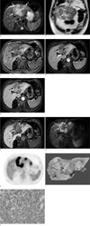 | Fig. 1A 78-year-old man with primary hepatic lymphoma. (a) Axial fat suppressed T2-weighted fast spin echo image shows a large lobulated hyperintense mass (arrows) in the left hepatic lobe. (b) Coronal T2-weighted single shot fast spin echo image shows a large lobulated heterogeneous hyperintense mass (arrows), surrounding the fissure for ligamentum teres (arrowhead). (c) Axial pre-contrast T1-weighted image shows hypointense mass (arrow) in the left hepatic lobe. (d, e) Axial portal venous (d) and equilibrium (e) phase contrast-enhanced T1-weighted image show poor, heterogeneous enhancement of the mass (arrow). Progressive enhancement is seen in the central portion of the mass. (f) Axial 20 minute-delayed hepatobiliary phase contrast-enhanced T1-weighted image clearly shows two different signal intensity of the lesion: lymphoma with very low signal intensity area (arrow) and slightly low signal intensity entrapped by the tumor with necrotic hepatic parenchyma (dotted arrow). (g) Axial diffusion-weighed image (b = 800) shows three different signal intensity of the lesion: a band-like high signal intensity of the tumor (arrow), thin linear dark signal intensity along the medial margin of the high signal intensity (arrowhead), consistent with necrotic tumor, and intermediate signal intensity area (dotted arrow) entrapped by the tumor consistent with necrotic hepatic parenchyma. (h) F-18 FDG PET-CT shows a band-like hot uptake showing the same configuration with that of diffusion-weighted image, suggestive of a viable tumor. (i) Gross specimen shows three different zones consisting of a band-like lobulated tumor (arrow), focal tumoral necrosis (arrowhead), and necrotic hepatic parenchyma without tumor involvement (dotted arrow). (j) Photomicrography of specimen (immunohistochemical staining for CD20, ×400) shows multiple large atypical lymphoid cells expressing CD20, consistent with diffuse large B-cell lymphoma.
|
References
1. Peixoto MCG, Filho AAA, Ribeiro ACR, D'lppolito G. Non-Hodgkin's lymphoma presenting as a single liver mass. Radiol Bras. 2009. 42:15–19.
2. Miyamoto Y, Izuo M, Ikeya T, et al. Right hepatic lobectomy for primary lymphoma: a case report and literature review. Jpn J Surg. 1986. 16:292–297.
3. Ryan J, Straus DJ, Lange C, et al. Primary lymphoma of the liver. Cancer. 1988. 61:370–375.
4. Noronha V, Shafi NQ, Obando JA, Kummar S. Primary non-Hodgkin's lymphoma of the liver. Crit Rev Oncol hematol. 2005. 53:199–207.
5. Maher MM, McDermott SR, Fenlon HM, et al. Imaging of primary non-Hodgkin's lymphoma of the liver. Clin Radiol. 2001. 56:295–301.
6. Gazelle GS, Lee MJ, Hahn PF, Goldberg MA, Rafaat N, Mueller PR. US, CT, and MRI of primary and secondary liver lymphoma. J Comput Assist Tomogr. 1994. 18:412–415.
7. Doi H, Horiike N, Hiraoka A, et al. Primary hepatic marginal zone B cell lymphoma of mucosa-associated lymphoid tissue type: case report and review of the literature. Int J Hematol. 2008. 88:418–423.
8. Rosai J. Rosai and Ackerman's surgical pathology. 9th ed. 1946–1947.
 XML Download