Journal List > Korean J Lab Med > v.31(2) > 1011728

Myoung, Shin, Lee, Kim, Shin, Suh, and Ryang: Multilocus Sequence Typing for Candida albicans Isolates from Candidemic Patients: Comparison with Southern Blot Hybridization and Pulsed-field Gel Electrophoresis Analysis

Abstract

Background

We evaluated the efficacy of multilocus sequence typing (MLST) for assessing the genetic relationship among Candida albicans isolates from patients with candidemia in a hospital setting.

Methods

A total of 45 C. albicans isolates from 21 patients with candidemia were analyzed. The MLST results were compared with results obtained by Southern blot hybridization (C1 fingerprinting) and pulsed-field gel electrophoresis (PFGE). PFGE analysis included karyotyping and restriction endonuclease analysis of genomic DNAs using BssHII (REAG-B) and SfiI (REAG-S).

Results

The 45 isolates yielded 20 unique diploid sequence types (DSTs) by MLST, as well as 12 karyotypes, 15 REAG-B patterns, 13 REAG-S patterns, and 14 C1 fingerprinting types. Microevolution among intra-individual isolates was detected in 6, 5, 3, 5, and 7 sets of isolates by MLST (1 or 2 allelic differences), REAG-B, REAG-S, C1 fingerprinting, and a combination of all methods, respectively. Among 20 DSTs, 17 were unique, and 3 were found in more than 1 patient. The results of 2 DSTs obtained from 9 patient isolates were in agreement with REAG and C1 fingerprinting patterns. However, the remaining DST, which was shared by 2 patient isolates, showed 2 different PFGE and C1 fingerprinting patterns. In addition, 3 sets of isolates from different patients, which differed in only 1 or 2 alleles by MLST, also exhibited different PFGE or C1 fingerprinting patterns.

Conclusions

MLST is highly discriminating among C. albicans isolates, but it may have some limitations in typing isolates from different patients, which may necessitate additional analysis using other techniques.

INTRODUCTION

Candida albicans is the most common species causing Candida bloodstream infections (BSI). Nosocomial C. albicans BSI develop primarily as a consequence of endogenous colonization, but are often caused by a strain acquired in hospital settings [1, 2]. Therefore, to identify the source of BSI caused by C. albicans, a suitable strain typing method is essential. In recent years, genomic microevolution (also called microvariation) has been documented for multiple C. albicans isolates from blood and other anatomic sites in individual patients with Candida BSI [1, 3-6]. Pfaller et al. [5] demonstrated that C. albicans BSI strains that are endemic in some hospitals undergo microevolution in the hospital setting. Given the rapid and frequent occurrence of C. albicans microevolution, assessing genetic relatedness among C. albicans isolates from patients with BSI is particularly important for understanding and controlling nosocomial C. albicans BSI within hospitals.
Multilocus sequence typing (MLST), which is based on DNA sequence analysis of nucleotide polymorphisms within housekeeping gene fragments, is a reliable, objective, and highly discriminating molecular typing method for the characterization of clinical C. albicans isolates [6, 7]. In addition, MLST permits the exchange of molecular typing data via the internet, which facilitates both local and global epidemiologic studies [7, 8]. To date, simultaneous comparisons of pulsed-field gel electrophoresis (PFGE), Southern blot hybridization, and MLST methods for genotyping C. albicans isolates from BSI patients has not been performed.
By using the MLST method, we analyzed the genetic relationships among C. albicans isolates from blood cultures and other anatomical sites from 21 patients with candidemia admitted to the same hospital over a 3-yr period. The purpose of this study was to evaluate the usefulness of MLST relative to PFGE and Southern blot hybridization fingerprinting tools for detecting nosocomial clusters vs. microevolution among C. albicans isolates from patients with C. albicans BSI.

MATERIALS AND METHODS

1. Patients and microorganisms

A total of 45 C. albicans isolates from 21 patients with candidemia were selected for this study. All isolates were obtained at Chonnam National University Hospital between September 1999 and August 2002. Previously described C. albicans strains from 2 nosocomial clusters of central venous catheter-related BSI (patients 15-21) were included in this study [3]. A single blood isolate was obtained from 10 patients, but multiple isolates (2-7) were obtained from 11 patients. C. albicans isolates were collected from cultures of clinical specimens before, during, or after collecting the isolates that caused candidemia. The interval between C. albicans isolation from blood culture and another body sites ranged between 1 and 32 days. Overall, isolates were cultured from blood (25 isolates from 21 patients), catheter tips (9 isolates from 7 patients), respiratory specimens (6 isolates from 3 patients), tissues (3 isolates from 1 patient), and urine (2 isolates from 2 patients). Species identification was based on colony morphology on CHROMagar Candida (BBL, Becton Dickinson, Sparks, MD, USA) at 35℃, microscopic morphology on cornmeal-Tween 80 agar, and use of a commercial system (API 20C; bioMérieux, Marcy L'Etoile, France, or the Vitek 2 system; Vitek 2 ID-YST, bioMérieux). We used the C. albicans strain, ATCC 20098, as a control in this study.

2. Multilocus sequence typing

C. albicans isolates were genotyped using the original MLST scheme described previously by Bougnoux et al. [6]. The internal regions of 7 housekeeping genes (AAT1a, ACC1, ADP1, MPIb, SYA1, VPS13, and ZWF1b) were sequenced. Sequence analysis was performed on both strands using an ABI Prism 3130xl Genetic Analyzer (Applied Biosystems, Foster City, CA, USA). Each strain was characterized by diploid sequence types (DSTs) resulting from the combination of genotypes obtained at 7 loci. Each strain was introduced into the original MLST online database (http://calbicans.mlst.net), which allowed us to compare these strains to those previously genotyped by this method and stored in the database.

3. Pulsed-field gel electrophoresis analysis

PFGE genotyping consisted of electrophoretic karyotyping and restriction endonuclease analysis of genomic DNA (REAG) using BssHII (REAG-B) and SfiI (REAG-S). PFGE methods for DNA preparation, REAG digestion, and electrophoresis have been presented in detail previously [3]. Isolates that differed by 1 or more bands were considered to have different karyotypes. To analyze the REAG patterns, isolates differing by 1 or more bands were designated as being different types. However, the strains with banding patterns that differed by 3 or fewer bands were described as subtypes (a or b) of a given clonal type. Serial isolates from the same patient showing a minor genotypic change (subtype) in the REAG pattern were said to have undergone microevolution.

4. Southern hybridization using the C1 fragment of the Ca3 probe

Southern blot hybridization using the 0.98-kb C1 fragment of Ca3 as a probe (C1 fingerprinting) was performed as previously described [3, 4]. Briefly, cells from single clonal colonies were transferred to YPD broth (1% yeast extract, 2% Bacto Peptone, 2% glucose; Difco, Detroit, MI, USA) and grown to the late log phase. DNA from each isolate was prepared using the method of Scherer and Stevens [9], digested with EcoRI, and separated on 0.8% agarose gels. Gels were stained with ethidium bromide to assess loading and transferred to nitrocellulose membranes. DNA fragments on the membranes were then hybridized with the radiolabeled C1 probe, as previously described [3, 4]. Blots were exposed to XAR-S film (Eastman Kodak Co., Rochester, NY, USA). Autoradiographs were examined visually to assess both band position and band intensity. Isolates determined to differ by 1 or more bands were designated as being a different type. C. albicans isolates from the same patient or the same clone showed genetic variation (i.e., 1 or 2 band differences), suggestive of microevolution [3, 4].

RESULTS

1. Sequence variability by MLST

A total of 2,883 bp from 7 housekeeping genes (AAT1a, ACC1, ADP1, MPIb, SYA1, VPS13, and ZWF1b) were sequenced in each of the 45 isolates from 21 patients with candidemia. Sixty-nine (2.4%) nucleotide sites were polymorphic (Table 1). There were 7 polymorphic sites per gene in the AAT1a and ACC1 loci, 9 in the SYA1 locus, 10 in the ZWF1b locus, 11 in the ADP1 locus, 12 in the VPS13 locus, and 13 in the MPIb locus. Thirty-one (44.9%) of the 69 individual changes were non-synonymous. The 7 housekeeping gene fragments sequenced displayed 8-10 different genotypes. The ACC1 (1), ADP1 (1), and SYA1 (2) fragments generated 4 new genotypes.
A total of 20 unique DSTs were obtained from a combination of genotypes from 7 housekeeping genes for the 45 clinical isolates. Twenty-three isolates from 14 patients belonged to previously described types (DSTs 90, 365, 656, 659, 601, 602, 693, 1395, 1646, and 1711), whereas 22 isolates from 12 patients belonged to new sequence types. Ten DSTs (DSTs 1788 to 1797) from our Korean strain collection novel to the internet DST database were added to the MLST database (http://calbicans.mlst.net).

2. Comparison of isolates from the same patient

In Table 2, the sequence of isolates, their anatomic sites, the date of isolation, and PFGE, C1 fingerprinting, and MLST types are presented for 45 isolates from each of the 21 patients. Two or more C. albicans isolates from blood cultures and other anatomic sites were obtained for 11 patients (patients 1-7, 9, 11, 16, and 18). In each of the 11 candidemic patients, C. albicans recovered from other sites and C. albicans recovered from blood showed identical or similar MLST, PFGE, and C1 fingerprinting types, except for the respiratory isolates from patient 2. Two respiratory isolates (isolates 2-3 and 2-4) from patient 2 showed different genotype patterns from blood isolates by all methods (Fig. 1).
When serial isolates from the same patient were examined using a combination of MLST, PFGE, and C1 fingerprinting methods, a total of 7 sets of intra-individual isolates showed microevolution. Catheter isolates from 4 patients (patients 1, 2, 9, and 18) and respiratory isolates from 2 patients (patients 6 and 11) exhibited microevolution relative to blood isolates. In addition, respiratory isolates from 1 patient (isolates 2-3 and 2-4) had also undergone microevolution. Of 7 sets of isolates, 6 gave non-identical sequencing results for 1 or 2 of the test allele loci analyzed with MLST. Each of these 7 sets of isolates had the same karyotype. Microevolution among intra-individual isolates was detected by REAG-B (5 sets), REAG-S (2 sets), and C1 fingerprinting (5 sets).

3. Comparison among isolates from different patients

PFGE analysis revealed 12 different karyotypes, 15 different REAG-B patterns, and 13 different REAG-S patterns among the isolates, whereas C1 fingerprinting identified 14 different types (Table 2). Of the 20 DSTs, 17 were unique for each patient, whereas 3 were found in more than 1 patient. Of 3 DSTs, 2 (DSTs 1711 and 1792) were shared by isolates from a previously well-characterized set of 2 nosocomial clusters. These 2 DSTs were shared by isolates from 9 pediatric patients (patients 13 to 21) admitted to the same intensive care unit. MLST results of blood isolates from these 9 patients were exactly matched by results from both PFGE (karyotyping, REAG-B, and REAG-S) and C1 fingerprinting. The remaining DST (DST 365) was shared by isolates from 2 different patients (4 isolates from patient 7 and isolate 2-4 from patient 2). Even though they were assigned to DST 365, each patient's isolates showed different PFGE and C1 fingerprinting patterns.
When the MLST sequence results from different patients were examined, 3 sets of isolates were found to have only 1 or 2 allelic differences (closely related). Of 3 sets, 1 set of isolates (isolate 8-1 and the isolates from patient 7) had similar C1 fingerprinting patterns, but different karyotyping and REAG patterns. In addition, 2 other sets of isolates (isolates 8-1 and 2-4; isolate 2-3 and the isolates from patient 7) exhibited different PFGE and C1 fingerprinting patterns. This result is in contrast to the finding that 2 respiratory isolates from the same patient (isolate 2-3 and isolate 2-4) showed 2 allelic differences (ACC1 and ADP1 genes) by MLST, but had very similar PFGE and C1 fingerprinting patterns (Fig. 1).

DISCUSSION

MLST allows the exchange of genotyping data via the internet for global epidemiology [7, 8]. A large amount of MLST data of C. albicans has accumulated from diverse locations, and it has been analyzed on a website. To date, an MLST study of C. albicans isolates has not been performed in Korea. The present study showed that some 
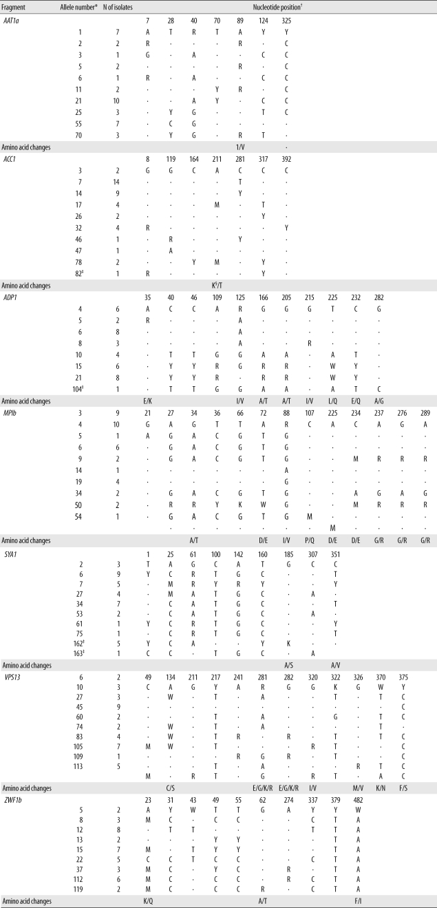
C. albicans strains that cause candidemia at our hospital shared the same MLST type with strains from other geographic regions of the world. Overall, 22 isolates from 12 patients belonged to new sequence types (10 DSTs), whereas 23 isolates from 14 patients belonged to previously described types (10 DSTs). In this study, 4 polymorphic nucleotide sites were identified. Our study showed that MLST allows an exchange of international genotyping data and offers distinctive standardization and portability.
Chen et al. [8] reported that MLST is superior to REAG-B for tracing the microevolution of 
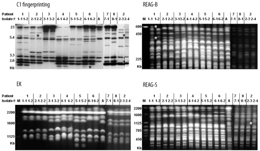
C. albicans strains within the same patient. However, simultaneous comparison of MLST, REAG-B, REAG-S, and C1 fingerprinting techniques for the detection of microevolution of C. albicans strains from patients with BSI has not been performed. In this study, multiple isolates from the same patient showing different DSTs were examined. By MLST, 1 or 2 allelic differences, indicative of microevolution [7], were found in 6 sets of isolates. REAG-B, REAG-S, and C1 fingerprinting also detected microevolution among 5, 2, and 5 sets of isolates from the same patient. However, none of these 4 methods detected all 7 sets of microevolution that were detected by combining all 4 methods used in this study. These results show that MLST is as efficient as C1 fingerprinting and REAG for detecting microevolution among sequential C. albicans isolates from Candida BSI.
Two previous studies showed that MLST analysis has the highest levels of concordance with Ca3 Southern hybridization [10] and REAG-B in discriminating epidemiologically related strains [
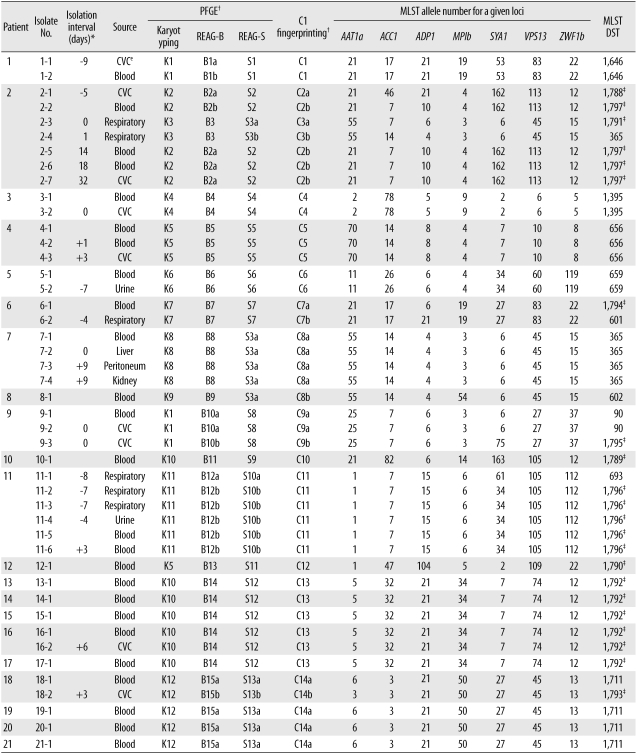
8]. We analyzed 2 previously recognized nosocomial clusters of BSI that had been evaluated by PFGE and C1 fingerprinting techniques with MLST. MLST analysis clearly identified 9 patients' isolates from 2 nosocomial clusters as 2 DSTs (DSTs 1711 and 1792). Of these 9 patient isolates, 1 catheter isolate from patient 18 showed a different DST (DST 1793) from corresponding blood isolates (DST 1711); however, it differed by only 1 locus, suggesting microevolution. This finding, together with microevolution observed among serial isolates from the same patient, suggest that closely related C. albicans strains, which have different DSTs only at 1 or 2 of the sequenced alleles, may be classified as epidemiologically related strains.
Of particular note in our study is that 1 set of isolates classified as the same DST (DST 365) by MLST differed in PFGE and C1 fingerprinting patterns. In addition, 3 sets of isolates from different patients in whom MLST showed 1 or 2 allelic differences (closely related), also revealed different patterns by PFGE or C1 fingerprinting. These isolates from 4 sets, which showed the same or similar MLST but exhibited different PFGE or C1 fingerprinting patterns, were obtained from 3 epidemiologically unrelated patients. These results suggest that MLST may be less useful for long-term epidemiological surveillance of C. albicans isolates in a hospital setting, and that REAG-B perhaps constitutes the more useful method for confirming MLST findings. In addition, our data show that MLST analysis of C. albicans isolates, in the absence of epidemiologic data, may not be recommended as a routine component of infection control, but that it may be useful in evaluating putative outbreaks identified by conventional hospital surveillance.
Odds et al. [11] also noted a few examples among their isolates that were very closely related according to MLST analysis, but that differed in ABC type, which is a very stable epidemiological marker in C. albicans isolates. Our results, together with those by Odds et al. [11], highlight the limitation of the MLST analysis in typing diverse strains of C. albicans from different patients (inter-patient analysis). These data suggest that frequent genetic evolutionary changes in C. albicans isolates may affect the interpretation of MLST data, especially for detecting inter- and intra-hospital spread of C. albicans strains. Therefore, the C. albicans strains at our hospital that shared the same DST with strains from other geographic regions of the world need further characterizing by PFGE or C1 fingerprinting.
In summary, we compared MLST, PFGE, and C1 fingerprinting for typing C. albicans isolates from patients with BSI. The results show that although MLST is a powerful tool for discriminating and detecting microevolution among C. albicans isolates, it has some limitations in typing diverse strains of C. albicans from different patients. Therefore, we suggest that REAG or C1 fingerprinting can be a very helpful confirmatory tool in instances when C. albicans isolates from different patients show the same or similar (differ by only 1 or 2 alleles) MLST types.

Acknowledgement

This work was partly supported by the Korea Research Fou-ndation Grant funded by the Korean Government (MO-EHRD) (KRF-2008-313-E00510).

Notes

No potential conflict of interest relevant to this article was reported.

References

1. Marco F, Lockhart SR, Pfaller MA, Pujol C, Rangel-Frausto MS, Wiblin T, et al. Elucidating the origins of nosocomial infections with Candida albicans by DNA fingerprinting with the complex probe Ca3. J Clin Microbiol. 1999; 37:2817–2828. PMID: 10449459.
2. Asmundsdóttir LR, Erlendsdóttir H, Haraldsson G, Guo H, Xu J, Gottfredsson M. Molecular epidemiology of candidemia: evidence of clusters of smoldering nosocomial infections. Clin Infect Dis. 2008; 47:e17–e24. PMID: 18549311.
crossref
3. Shin JH, Park MR, Song JW, Shin DH, Jung SI, Cho D, et al. Microevolution of Candida albicans strains during catheter-related candidemia. J Clin Microbiol. 2004; 42:4025–4031. PMID: 15364985.
4. Lockhart SR, Fritch JJ, Meier AS, Schröppel K, Srikantha T, Galask R, et al. Colonizing populations of Candida albicans are clonal in origin but undergo microevolution through C1 fragment reorganization as demonstrated by DNA fingerprinting and C1 sequencing. J Clin Microbiol. 1995; 33:1501–1509. PMID: 7650175.
5. Pfaller MA, Lockhart SR, Pujol C, Swails-Wenger JA, Messer SA, Edmond MB, et al. Hospital specificity, region specificity, and fluconazole resistance of Candida albicans bloodstream isolates. J Clin Microbiol. 1998; 36:1518–1529. PMID: 9620370.
6. Bougnoux ME, Tavanti A, Bouchier C, Gow NA, Magnier A, Davidson AD, et al. Collaborative consensus for optimized multilocus sequence typing of Candida albicans. J Clin Microbiol. 2003; 41:5265–5266. PMID: 14605179.
7. Bougnoux ME, Aanensen DM, Morand S, Théraud M, Spratt BG, d'Enfert C. Multilocus sequence typing of Candida albicans: strategies, data exchange and applications. Infect Genet Evol. 2004; 4:243–252. PMID: 15450203.
8. Chen KW, Chen YC, Lo HJ, Odds FC, Wang TH, Lin CY, et al. Multilocus sequence typing for analyses of clonality of Candida albicans strains in Taiwan. J Clin Microbiol. 2006; 44:2172–2178. PMID: 16757617.
9. Scherer S, Stevens DA. Application of DNA typing methods to epidemiology and taxonomy of Candida species. J Clin Microbiol. 1987; 25:675–679. PMID: 3033016.
10. Chowdhary A, Lee-Yang W, Lasker BA, Brandt ME, Warnock DW, Arthington-Skaggs BA. Comparison of multilocus sequence typing and Ca3 fingerprinting for molecular subtyping epidemiologically-related clinical isolates of Candida albicans. Med Mycol. 2006; 44:405–417. PMID: 16882607.
11. Odds FC, Bougnoux ME, Shaw DJ, Bain JM, Davidson AD, Diogo D, et al. Molecular phylogenetics of Candida albicans. Eukaryot Cell. 2007; 6:1041–1052. PMID: 17416899.
Fig. 1
Representative genotyping patterns of Candida albicans obtained by Southern hybridization with the C1 fragment of the Ca3 probe (C1 fingerprinting), electrophoretic karyotyping (EK), and restriction endonuclease analysis of genomic DNA using SfiI (REAG-S) and BssHII (REAG-B). See Table 2 for detailed information on each isolate from 8 patients (patients 1-8). All sequential isolates had the same karyotype and showed the same or similar REAG and C1 fingerprinting patterns, except for 2 isolates (2-3 and 2-4) from patient 2. Stars indicate the positions of added or deleted bands in the minor patterns for clonal strains from the same patient, suggesting that microevolution had occurred. Three sets of isolates from different patients (isolates 7-1 and 8-1; isolates 8-1 and 2-4; isolates 7-1 and 2-3), which differed in only 1 or 2 alleles according to MLST analysis, revealed different PFGE or C1 fingerprinting patterns.
Abbreviations: A, C. albicans ATCC 90028; M, Saccharomyces cerevisiae DNA concatamers as a molecular size marker.
kjlm-31-107-g001
Table 1
Positions of polymorphic nucleotide sites, associated amino acid changes, and allele number at the 7 loci used for multilocus sequence typing analysis
kjlm-31-107-i001

*Allele numbers are based on those in the MLST database (http://test1.mlst.net); One letter IUPAC nucleotide code: R=A or G; M=A or C; W=A or T; Y=C or T; K=G or T; S=C or G; Indicates new (unpublished) genotypes identified in this study; §Single letter amino acid symbols.

Table 2
Genotyping results for Candida albicans from blood and other body sites from 21 patients with candidemia
kjlm-31-107-i002

*Interval between the isolation of C. albicans from a blood culture and other body sites in each patient, which is presented as the number of days before (-) or after (+) the first positive blood culture; The differences denoted, a or b, are insufficient to classify the isolate as different rather than similar (sharing all of the bands except 1 or 2 bands); Indicates new (unpublished) diploid sequence types identified in this study.

Abbreviations: PFGE, pulsed-field gel electrophoresis; REAG, restriction endonuclease analysis of genomic DNA; MLST, multilocus sequence typing; DST, diploid sequence type; CVC, central venous catheter.

TOOLS
Similar articles