Abstract
Hypermethylation of the homeobox (HOX) gene promoter leads to decreased expression of the gene during tumor development and is thought to be correlated with the clinical outcome in leukemia. In this study, we performed pyrosequencing to quantify the methylation level of HOXA5 genes in the bone marrow samples obtained from 50 patients with AML and 19 normal controls. The methylation percentage of HOXA5 in AML patients (median=65.4%, interquartile range=35.9-72.3%) was higher than that of HOXA5 in control patients (median=43.1%, interquartile range=36.7-49.6%, Mann-Whitney U test, P=0.012). The patients of the AML group who had a high methylation percentage (>70%) had a good prognosis with a 3-yr overall survival (OS) of 82.5%, whereas the patients with a low methylation percentage (≤70%) showed a 3-yr OS of 40.5% (P=0.048). Cox proportional hazards regression showed that the methylation percentages of HOXA5 were independently associated with the 3-yr OS of AML patients, regardless of their karyotypes. We propose that the quantification of HOXA5 methylation by pyrosequencing may be useful for predicting short-term prognosis in AML. However, the limitations of our study are the small sample size and its preliminary nature. Thus, a larger study should be performed to clearly determine the relationships among HOXA5 methylation levels, cytogenetics, and prognosis in AML patients.
Go to : 
REFERENCES
1.Cillo C., Cantile M., Faiella A., Boncinelli E. Homeobox genes in normal and malignant cells. J Cell Physiol. 2001. 188:161–9.

2.Abramovich C., Humphries RK. Hox regulation of normal and leukemic hematopoietic stem cells. Curr Opin Hematol. 2005. 12:210–6.
3.Bird AP., Wolffe AP. Methylation-induced repression—belts, braces, and chromatin. Cell. 1999. 99:451–4.
4.Herman JG., Baylin SB. Gene silencing in cancer in association with promoter hypermethylation. N Engl J Med. 2003. 349:2042–54.

5.Strathdee G., Sim A., Parker A., Oscier D., Brown R. Promoter hypermethylation silences expression of the HoxA4 gene and correlates with IgVh mutational status in CLL. Leukemia. 2006. 20:1326–9.
6.Strathdee G., Holyoake TL., Sim A., Parker A., Oscier DG., Melo JV, et al. Inactivation of HOXA genes by hypermethylation in myeloid and lymphoid malignancy is frequent and associated with poor prognosis. Clin Cancer Res. 2007. 13:5048–55.
7.Drabkin HA., Parsy C., Ferguson K., Guilhot F., Lacotte L., Roy L, et al. Quantitative HOX expression in chromosomally defined subsets of acute myelogenous leukemia. Leukemia. 2002. 16:186–95.
8.Golub TR., Slonim DK., Tamayo P., Huard C., Gaasenbeek M., Mesirov JP, et al. Molecular classification of cancer: class discovery and class prediction by gene expression monitoring. Science. 1999. 286:531–7.

9.Grimwade D., Walker H., Oliver F., Wheatley K., Harrison C., Harrison G, et al. The importance of diagnostic cytogenetics on outcome in AML: analysis of 1,612 patients entered into the MRC AML 10 trial. The Medical Research Council Adult and Children's Leukaemia Working Parties. Blood. 1998. 92:2322–33.
10.Strathdee G., Sim A., Soutar R., Holyoake TL., Brown R. HOXA5 is targeted by cell-type-specific CpG island methylation in normal cells and during the development of acute myeloid leukaemia. Carcinogenesis. 2007. 28:299–309.
11.Vieille-Grosjean I., Roullot V., Courtois G. Identification of homeobox-containing genes expressed in hematopoietic blast cells. Biochem Biophys Res Commun. 1992. 185:785–92.

12.Moretti P., Simmons P., Thomas P., Haylock D., Rathjen P., Vadas M, et al. Identification of homeobox genes expressed in human haemopoietic progenitor cells. Gene. 1994. 144:213–9.

13.Andreeff M., Ruvolo V., Gadgil S., Zeng C., Coombes K., Chen W, et al. HOX expression patterns identify a common signature for favorable AML. Leukemia. 2008. 22:2041–7.
14.Debernardi S., Lillington DM., Chaplin T., Tomlinson S., Amess J., Rohatiner A, et al. Genome-wide analysis of acute myeloid leukemia with normal karyotype reveals a unique pattern of homeobox gene expression distinct from those with translocation-mediated fusion events. Genes Chromosomes Cancer. 2003. 37:149–58.

15.Celetti A., Barba P., Cillo C., Rotoli B., Boncinelli E., Magli MC. Characteristic patterns of HOX gene expression in different types of human leukemia. Int J Cancer. 1993. 53:237–44.
Go to : 
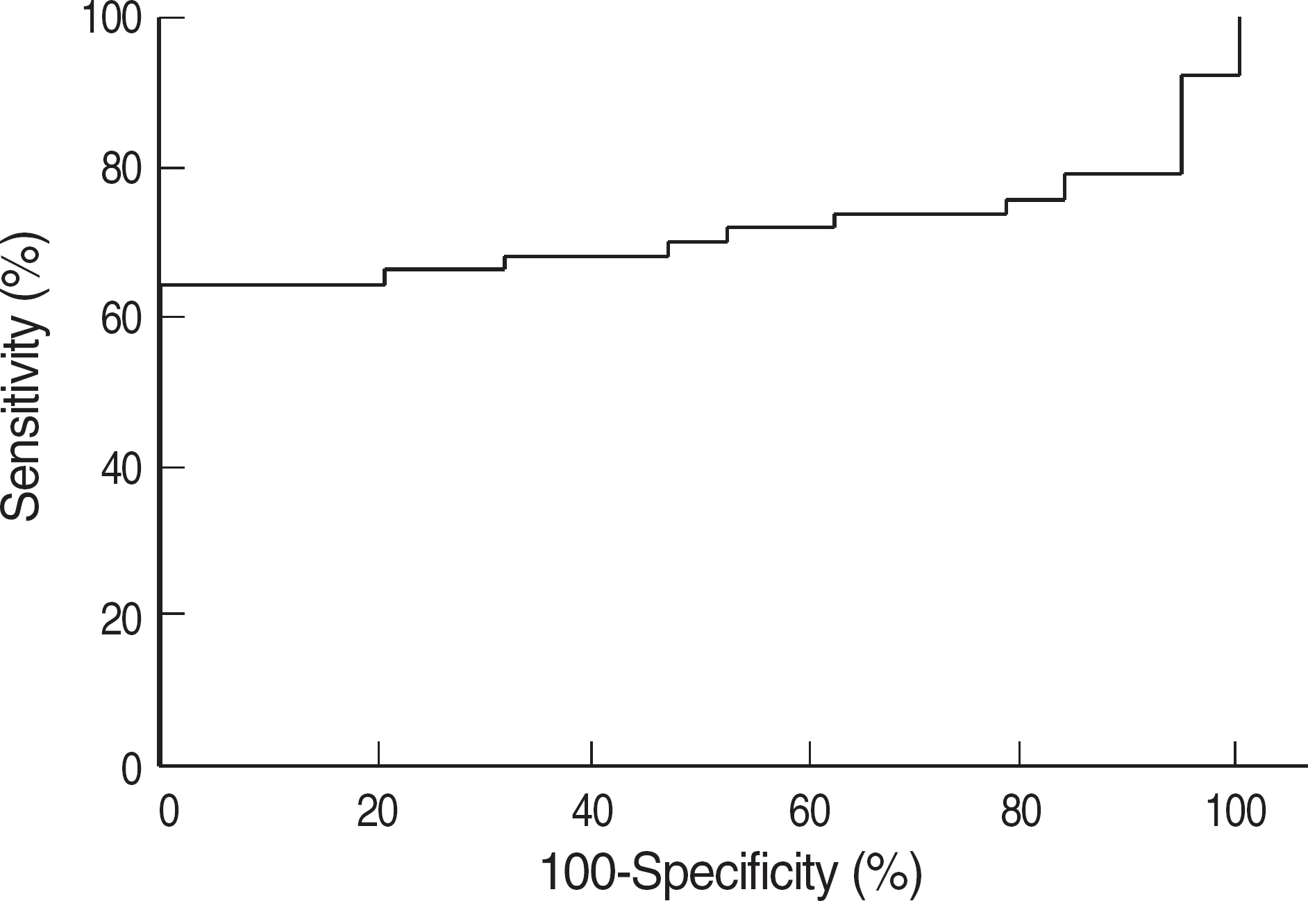
 | Fig. 1.ROC curve analysis showed that a cut-off of 70% HOXA5 methylation yielded sensitivity and specificity of 60% and 100%, respectively. |
 | Fig. 2.Percentage of 3-yr overall survival of patients with “mean HOXA5 methylation percentage greater than 70%” and of those with “mean HOXA5 methylation less than 70%”. |
Table 1.
Characteristics of AML patients according to HOXA5 methylation
| Mean of MtP | P value | ||
|---|---|---|---|
| Less than 70% (N=32) | More than 70% (N=18) | ||
| Age (mean±SD) | 51.9±14.2 | 47.1±16.5 | 0.201 |
| Sex (%) | |||
| Male | 15 (46.9) | 13 (72.2) | |
| Female | 17 (53.1) | 5 (27.8) | |
| FAB classification (%) | 0.452 | ||
| AML, unclassified | 3 (9.3) | 0 (0.0) | |
| M0 | 2 (6.3) | 0 (0.0) | |
| M1 | 7 (21.9) | 7 (38.9) | |
| M2 | 9 (28.1) | 3 (16.7) | |
| M3 | 4 (12.5) | 6 (33.3) | |
| M4 | 5 (15.6) | 1 (5.6) | |
| M5 | 1 (3.1) | 1 (5.6) | |
| M6 | 1 (3.1) | 0 (0.0) | |
| Karyotyping (%) | 0.004 | ||
| Unfavorable | 4 (12.5) | 3 (17.6) | |
| Intermediate | 24 (75.0) | 5 (29.4) | |
| Favorable | 4 (12.5) | 9 (52.9) | |
| Normal karyotype | 17 (70.8) | 4 (80.0) | 1.000 |
| (%) among | |||
| intermediate group | |||
| Chemotherapy | 0.114 | ||
| regimens (%) | |||
| AI | 25 (78.1) | 9 (50.0) | |
| AIDA | 4 (12.5) | 6 (33.3) | |
| Other | 3 (9.4) | 3 (16.7) | |
| LDH (mean±SD)∗ | 1,597.5±1,464.6 1,458.9±1,505.3 | 0.303 | |
| Hb (mean±SD)∗ | 8.5±1.5 | 8.5±1.2 | 0.973 |
| WBC (×103/μL, mean±SD)∗ | 29.5±60.6 | 38.0±67.7 | 0.412 |
Table 2.
Cox proportional hazard regression analysis for 3-yr overall survival of AML patients


 PDF
PDF ePub
ePub Citation
 Print
Print


 XML Download