

 PDF
PDF ePub
ePub Citation
 Print
Print


Abstract
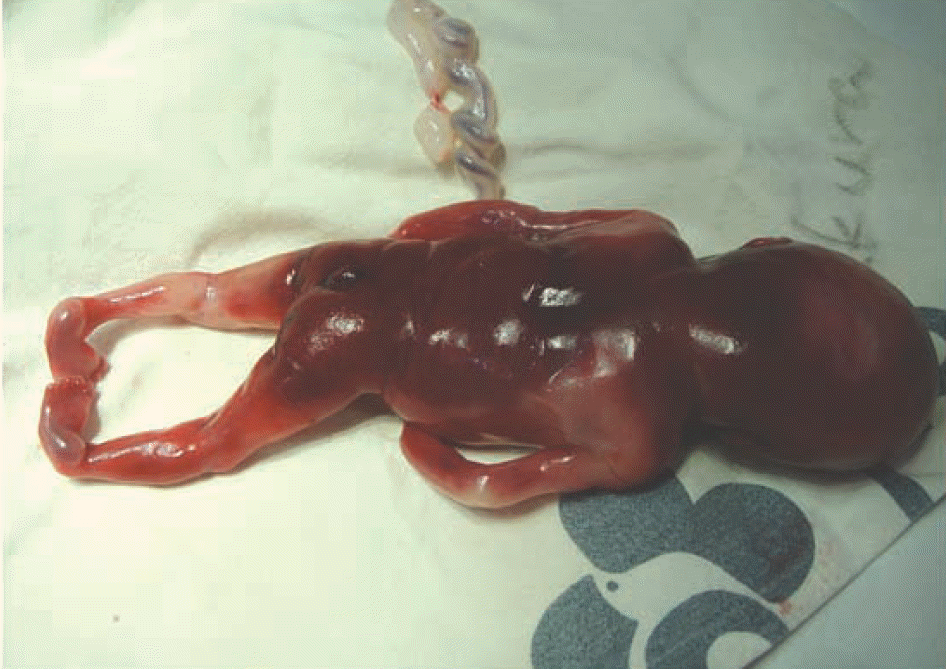
Although trisomy 18 (Edwards' syndrome) or the terminal deletion syndromes of 18p and 18q have been occasionally detected, pseudoisodicentric chromosome 18 is a very rare constitutional chromosomal abnormality. We describe a case of pseudoisodicentric chromosome 18q without mosaicism, which was confirmed from fetal cells in the amniotic fluid used for prenatal diagnosis of multiple congenital anomalies. A 23-yr-old pregnant woman was suspected of having a fetal anomaly at 18+3 weeks gestation. In sonography, the fetus showed multiple anomalies: bilateral overt ventriculomegaly in the brain, ventricular septal defect and valve anomaly in the heart, bilateral club foot, polydactyly, meningocele, and a single umbilical artery. The pregnancy was terminated and a conventional G-banded chromosome study was performed using amniotic fluid. Twenty metaphase cells among the cultured amniocytes showed a 46,XX,psu idic(18)(q22). Consequently, the fetus had partial trisomy (18pter→q22) and partial monosomy (18q22→qter). Both parents were confirmed to have a normal karyotype.
Go to : 
REFERENCES
1.Lin CC., Li YC., Liu PP., Hsieh LJ., Cheng YM., Teng RH, et al. Identification and characterization of a new type of asymmetrical dicentric chromosome derived from a single maternal chromosome 18. Cytogenet Genome Res. 2007. 119:291–6.

2.Morrissette JJ., Medne L., Bentley T., Owens NL., Geiger E., Pipan M, et al. A patient with mosaic partial trisomy 18 resulting from dicentric chromosome breakage. Am J Med Genet A. 2005. 137:208–12.
3.Ward BE., Bradley CM., Cooper JB., Robinson A. Homodicentric chromosomes: a distinctive type of dicentric chromosome. J Med Genet. 1981. 18:54–8.
4.Lemyre E., der Kaloustian VM., Duncan AM. Stable non-Robertsonian dicentric chromosomes: four new cases and a review. J Med Genet. 2001. 38:76–9.
5.Higgins AW., Gustashaw KM., Willard HF. Engineered human dicentric chromosomes show centromere plasticity. Chromosome Res. 2005. 13:745–62.
6.Therman E., Trunca C., Kuhn EM., Sarto GE. Dicentric chromosomes and the inactivation of the centromere. Hum Genet. 1986. 72:191–5.
7.Madan K., Vlasveld L., Barth PG. Ring-18 and isopseudodicentric-18 in the same child: a hypothesis to account for common origin. Ann Genet. 1981. 24:12–6.
8.Meins M., Böhm D., Grossmann A., Herting E., Fleckenstein B., Fauth C, et al. First non-mosaic case of isopseudodicentric chromosome 18 (psu idic(18)(pter → q22.1::q22.1 → pter) is associated with multiple congenital anomalies reminiscent of trisomy 18 and 18q- syndrome. Am J Med Genet A. 2004. 127A:58–64.
9.Brandt CA., Djernes B., Str⊘mkjaer H., Petersen MB., Pedersen S., Hindkjaer J, et al. Pseudodicentric chromosome 18 diagnosed by chromosome painting and primed in situ labelling (PRINS). J Med Genet. 1994. 31:99–102.
10.Matsuoka R., Matsuyama S., Yamamoto Y., Kuroki Y., Matsui I. Trisomy 18q. A case report and review of karyotype-phenotype correlations. Hum Genet. 1981. 57:78–82.
11.Oudesluijs GG., Hulzebos CV., Sikkema-Raddatz B., Van Essen AJ. Mosaic isodicentric chromosome 18q: sixth report and review. Genet Couns. 2006. 17:395–400.
12.Feenstra I., Vissers LE., Orsel M., van Kessel AG., Brunner HG., Veltman JA, et al. Genotype-phenotype mapping of chromosome 18q deletions by high-resolution array CGH: an update of the phenotypic map. Am J Med Genet A. 2007. 143A:1858–67.
13.Wallerstein R., Yu MT., Neu RL., Benn P., Lee Bowen C., Crandall B, et al. Common trisomy mosaicism diagnosed in amniocytes involving chromosomes 13, 18, 20 and 21: karyotype-phenotype correlations. Prenat Diagn. 2000. 20:103–22.
Go to :
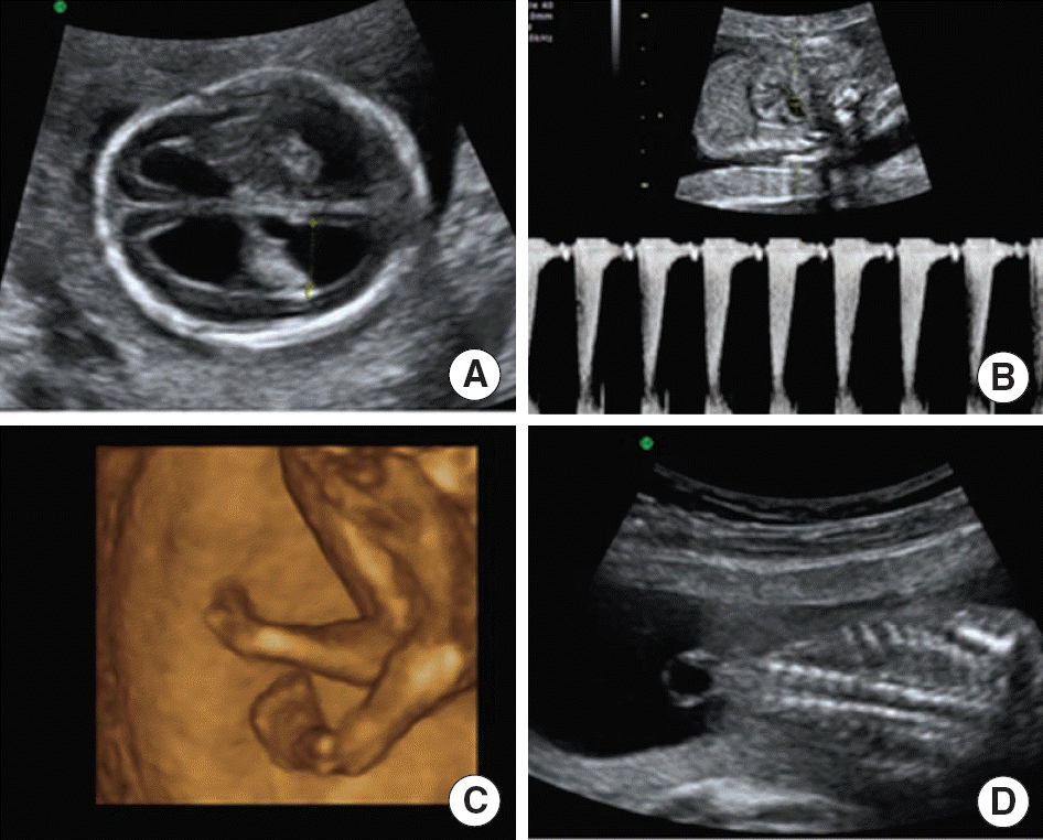 | Fig. 1.Prenatal sonographic findings of pseudoisodicentric 18q at 18+3 weeks gestation. (A) Transaxial plane of the fetal brain shows bilateral overt ventriculomegaly. (B) Fetal echocardiography shows pulmonary stenosis (2 m/s). (C) 3D ultrasonography shows bilateral club foot. (D) Coronal plane of the fetal spine shows meningocele. |
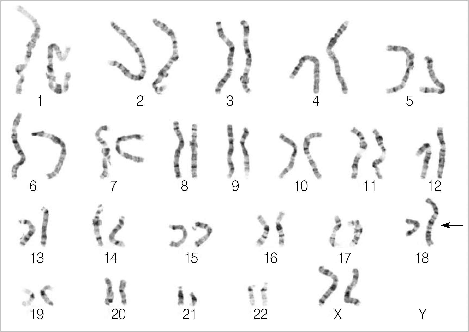 | Fig. 3.Karyotype showing pseudoisodicentric chromosome 18 by G-banding analysis. Chromosome 18 shows a breakpoint at 18q22 and has 2 centromeres, the upper one of which has a primary constriction, whereas the lower one lacks a primary constriction. |
Table 1.
Summary of clinical findings for the present case, compared with two other cases of psu idic 18q without mosaicism
| Cases of psu idic 18q without mosaicism | ||||
|---|---|---|---|---|
| Findings of pure trisomy 18 | 1st case [Meins et al.8] | 2nd case [Lin et al.1] | Present study [Cho et al.] | |
| 1. Karyotype | 47,XX,+18, or 47,XY,+18 | 46,XX,psu dic(18)(q22.1) | 46,XX,psu dic(18) | 46,XX,psu idic(18)(q22) |
| 2. Intrauterine growth retardation | Detected | Detected | NC | Detected |
| 3. Brain | Ventriculomegaly | Normal | Normal | Ventriculomegaly |
| 4. Face | Cleft lip and palate, micrognathia, small and low-set ears | Cleft lip and palate | Cleft lip and palate, anotia Rt. ear), low-set left ear, micrognathia | Normal |
| 5. Neck | No special anomaly | Short | NC | Normal |
| 6. Thorax | No special anomaly | Narrow | NC | Normal |
| 7. Heart | VSD, ASD, PDA | VSD and ASD | Normal | VSD and valve anomaly |
| 8. Kidney | Hydronephrosis, horseshoe kidney | Real hypoplasia | Normal | Normal |
| 9. Spinal cord | Neural tube defect | NC | NC | Meningocele |
| 10. Skin | Cutis laxa | Redundant skin | NC | Normal |
| 11. Extremities | Club feet, rocker-bottom feet | Club foot and hand, thumb aplasia | Rocker-bottom feet, abnormal finger∗ and toe† | Club feet and polydactyly |
| 12. Umbilical artery | Single umbilical artery | NC | NC | Single umbilical artery |
 XML Download