

 PDF
PDF ePub
ePub Citation
 Print
Print


Abstract
Background:
Blood glucose testing (BGT) at the forearm minimizes the pain experienced during sampling of capillary blood. We compared the BGT results for forearm sampling with those for standard finger skin puncture and venous serum to evaluate the clinical validity of forearm BGT.
Methods:
BGT was performed on the finger (GF) and forearm (GA) with a portable glucometer in 555 subjects, including 61 diabetic patients, under fasting conditions. BGT with venous serum (GV) was followed within an hour in 514 subjects. Simple linear regression, intraclass correlation, and Passing-Bablok regression analyses were performed using the GA-GF and GA-GV data.
Results:
GA showed an excellent linear relationship with both GF and GV with a Pearson correlation coefficient (r) of 0.97 (P<0.0001) in the patient group, which was similar to the findings in the normal group except for the lower r values. The mean bias between GA and GF and between GA and GV were within ±10 mg/dL in both groups. The intraclass correlation coefficients were slightly smaller than the corresponding r values, but they showed the same tendency in both groups. In the Passing-Bablok analyses, the 95% confidence intervals of the slope and intercept parameters were <± 20% of unity and <±20 mg/dL, respectively, which were within the acceptable ranges. All 3 statistical analyses supported the satisfactory agreement of GA with GF or GV.
Go to : 
REFERENCES
1.Zimmet P., Alberti KG., Shaw J. Global and societal implications of the diabetes epidemic. Nature. 2001. 414:782–7.

2.Kim MH., Kim MK., Choi BY., Shin YJ. Educational disparities in the metabolic syndrome in a rapidly changing society: the case of South Korea. Int J Epidemiol. 2005. 34:1266–73.
3.Cho NH. Diabetes epidemiology in Korean. J Korean Diabetes Assoc. 2001. 25:1–10.
4.Korea National Statistical Office. Annual report on the cause of death statistics (based on vital registration) 2006. Statstice Korea;2007.
5.Kim EJ, Min HK, editors. Diabetics. 3rd ed.Seoul: Korea Medical Book Company;2005. :1.
6.Parchman ML., Pugh JA., Noel PH., Larme AC. Continuity of care, self-management behaviors, and glucose control in patients with type 2 diabetes. Med Care. 2002. 40:137–44.
7.Kim EJ, Min HK, editors. Diabetics. 2nd ed.Seoul: Korea Medical Book Company;1998. p. 303–8.
8.Skyler JS. Self-monitoring of blood glucose. Med Clin North Am. 1982. 66:1227–50.
9.Choi JY. A comparison on the degree of pain according to methods of blood sugar test between DM patients and healthy group. J Korean Acad Nurs. 2003. 33:928–35.
10.Park SW., Kim DJ., Kim JY., Kim HY., Min KW., Baek SH, et al. Current status of diabetes management in Korea using national insurance database. Korean Diabetes Association 19th Spring Meeting. 2006. 227–8.
11.Cowett RM., D'Amico LB. Capillary (heelstick) versus venous blood sampling for the determination of glucose concentration in the neonate. Biol Neonate. 1992. 62:32–6.
12.Barker DP., Rutter N. Exposure to invasive procedures in neonatal intensive care unit admissions. Arch Dis Child Fetal Neonatal Ed. 1995. 72:F47–8.
13.McIntosh N., van Veen L., Brameyer H. Alleviation of the pain of heel prick in preterm infants. Arch Dis Child Fetal Neonatal Ed. 1994. 70:F177–81.
14.Cunningham DD., Henning TP., Shain EB., Young DF., Elstrom TA., Taylor EJ, et al. Vacuum-assisted lancing of the forearm: an effective and less painful approach to blood glucose monitoring. Diabetes Technol Ther. 2000. 2:541–8.
15.Park MS., Park KS., Kim KA., Jun MH., Kim TI., Lee TS, et al. Vacuum assisted auto-lancing technique for capillary blood sampling on the forearm with minimized pain. J Biomed Eng Res. 2004. 25:557–63.
16.Lock JP., Szuts EZ., Malomo KJ., Anagnostopoulos A. Whole-blood glucose testing at alternate sites: glucose values and hematocrit of capillary blood drawn from fingertip and forearm. Diabetes Care. 2002. 25:337–41.
17.Cohen M., Boyle E., Delaney C., Shaw J. A comparison of blood glucose meters in Australia. Diabetes Res Clin Pract. 2006. 71:113–8.
18.Reinauer H., Home RD, et alLaboratory diagnosis and monitoring of diabetes mellitus. Geneva, Switzerland: World Health Organization. 2002. 11–2.
19.U. S. Food and Drug Administration. Review criteria for assessment of portable blood glucose monitoring in-vitro diagnostic devices using glucose oxidase, dehydrogenase, or hexokinase methodology. Guidance document (medical devices). U. S. Department of Health and Human Service. 1997. 5.
20.Koch GG. Intraclass correlation coefficient. Kotz S, Johnson NL, editors. Encyclopedia of statistical sciences. New York: John Wiley & Sons;1982. p. 213–7.
21.Passing H., Bablok W. A new biometrical procedure for testing the equality of measurements from two different analytical methods. Application of linear regression procedures for method comparison studies in clinical chemistry, Part I. J Clin Chem Clin Biochem. 1983. 21:709–20.
22.National Committee for Clinical Laboratory Standards. Point-of-care blood glucose testing in acute and chronic care facilities: approved guideline. Document C30-A2. 2nd ed.Wayne, PA: NCCLS;2002. ;22.
23.International Organization for Standardization. ISO 15197:2003. In vitro diagnostic test systems requirements for blood-glucose monitoring systems for self-testing in managing diabetes mellitus. Geneva: International Organization for Standardization. 2003.
24.Clarke WL., Cox D., Gonder-Frederick LA., Carter W., Pohl SL. Evaluating clinical accuracy of systems for self-monitoring of blood glucose. Diabetes Care. 1987. 10:622–8.
25.Kim DK., Won JH., Potapov SN., Meriakri VV., Chigryai EE. Basic investigation for the non-invasive measurement of blood glucose concentrations by millimeter waves. IEEK Journal (SC). 1998. 42:3–6.
26.Waynant RW., Chenault VM. Overview of non-invasive fluid glucose measurement using optical techniques to maintain glucose control in diabetes mellitus. LEOS newsletter. 1998. 12:3–6.
27.Paul EK., Ruth SW., John BN. Carbohydrates. Henry JB, editor. Clinical diagnosis and management by laboratory methods. Philadelphia, PA: WB Sanders;2001. :214.
28.Kwon MJ., Lee SY. Evaluation of GLUCOCARD X-METER glucose monitoring system. Korean J Lab Med. 2008. 28:8–15.
29.Jungheim K., Koschinsky T. Glucose monitoring at the arm: risky delays of hypoglycemia and hyperglycemia detection. Diabetes Care. 2002. 25:956–60.
30.Jungheim K., Koschinsky T. Risky delay of hypoglycemia detection by glucose monitoring at the arm. Diabetes Care. 2001. 24:1303–6.
31.Ellison JM., Stegmann JM., Colner SL., Michael RH., Sharma MK., Ervin KR, et al. Rapid changes in postprandial blood glucose produce concentration differences at finger, forearm, and thigh sampling sites. Diabetes Care. 2002. 25:961–4.
32.Fagrell B. Microcirculation of the skin. In the physiology and pharmacology of the microcirculation. Orlando: Academic Press. 1984. 133–80.
33.Rendell M., Bergman T., O'Donnell G., Drobny E., Borgos J., Bonner RF. Microvascular blood flow, volume, and velocity measured by laser Doppler techniques in IDDM. Diabetes. 1989. 38:819–24.
34.Stansberry KB., Peppard HR., Babyak LM., Popp G., McNitt PM., Vinik AI. Primary nociceptive afferents mediate the blood flow dysfunction in non-glabrous (hairy) skin of type 2 diabetes: a new model for the pathogenesis of microvascular dysfunction. Diabetes Care. 1999. 22:1549–54.
35.McGarraugh G. Response to Jungheim and Koschinsky: glucose monitoring at the arm. Diabetes Care. 2001. 24:1304–6.
36.Pohl SL., Gonder-Frederick L., Cox DJ., Evans WS. Self-measurement of blood glucose concentration: clinical significance of patient-generated measurements. Diabetes Care. 1985. 8:617–9.
37.Tieszen KL., New JP. Alternate site blood glucose testing: do patients prefer it? Diabet Med. 2003. 20:325–8.
38.Dai KS., Tai DY., Ho P., Chen CC., Peng WC., Chen ST, et al. Accuracy of the EasyTouch blood glucose self-monitoring system: a study of 516 cases. Clin Chim Acta. 2004. 349:135–41.
Go to :
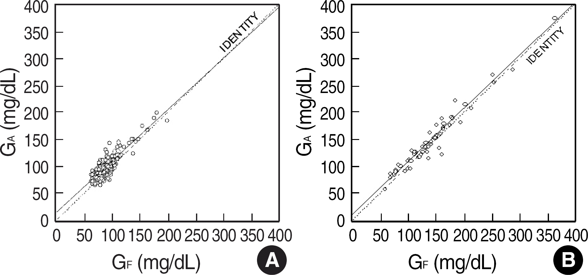 | Fig. 1.Simple linear regression results between the forearm (GA) and the finger (GF) glucose measurements in the normal (A) and the patient (B) groups. The solid and dotted lines represent the regression and the identity lines, respectively. |
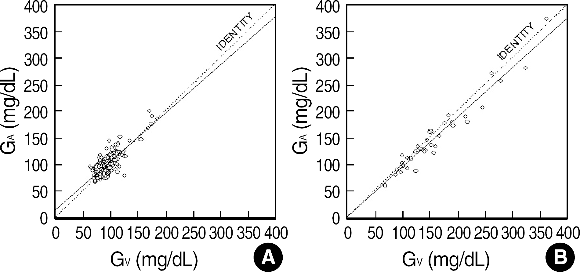 | Fig. 2.Simple linear regression results between the forearm (GA) and the vein (GV) glucose measurements in the normal (A) and the patient (B) groups. The solid and dotted lines represent the regression and the identity lines, respectively. |
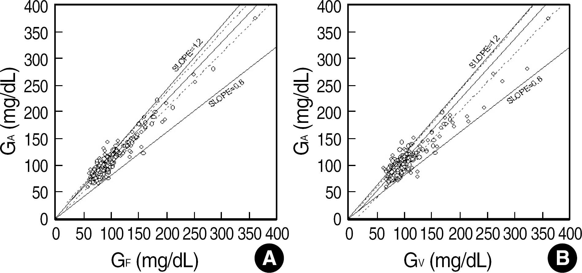 | Fig. 3.Results of the Passing-Bablok regression analysis between the forearm (GA) and the finger (GF) (A) and between the forearm and the vein (GV) (B) presented with the 95% confidence limit lines (dotted lines) and acceptable limit lines (solid lines with the slopes of 0.8 and 1.2). |
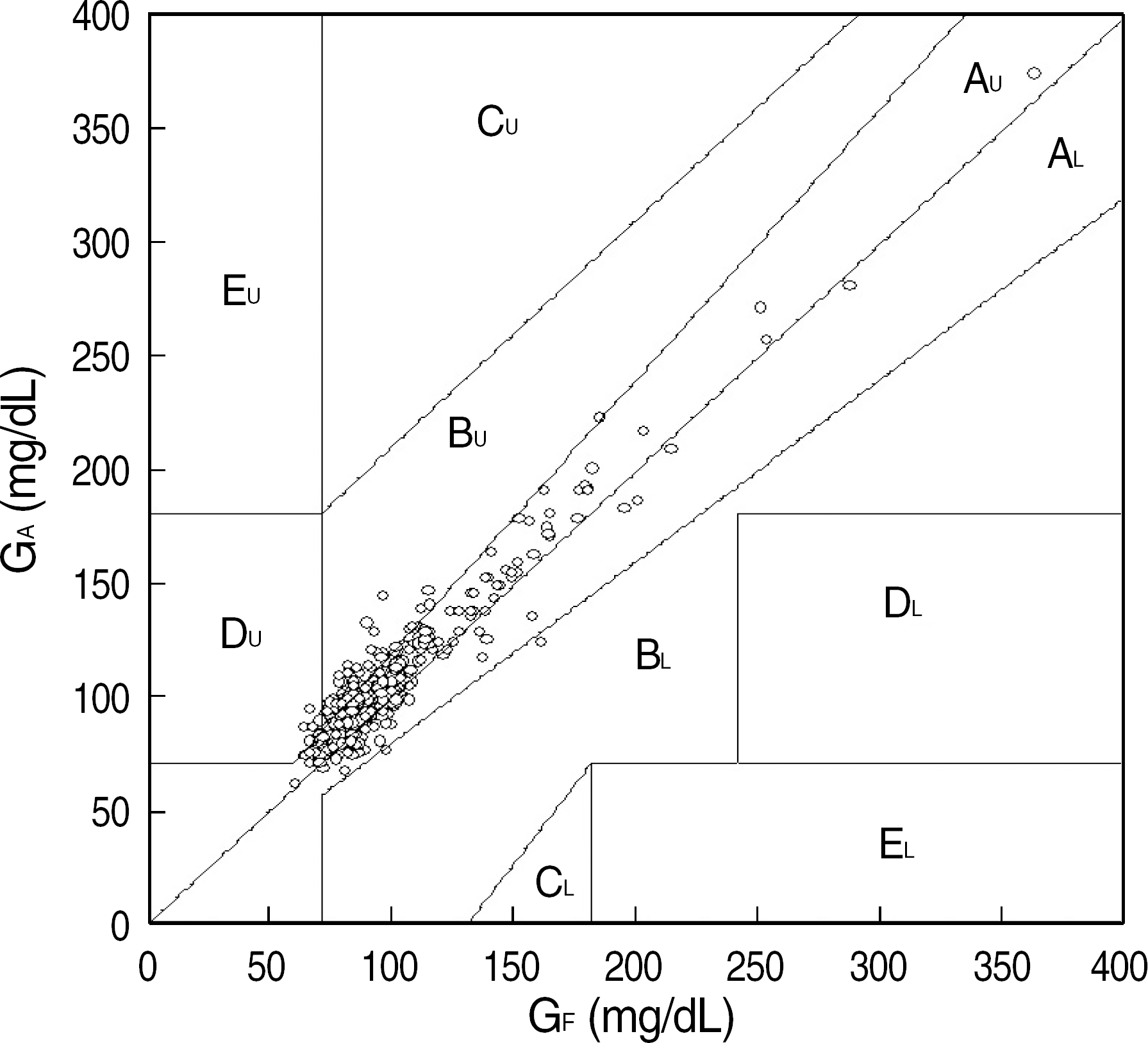 | Fig. 4.Individual blood glucose data points obtained from the forearm (GA) and finger (GF) plotted in the GA-GF plane superimposed on the error grid. In addition, the figure shows clinically accepted zones (denoted by AU,L and BU,L) in which 98% of the data are included (refer to text). |
Table 1.
General characteristics of the subjects
Table 2.
Mean (SD) values of the differences in glucose measurements in the normal and the patient groups
| Normal | Patient | |
|---|---|---|
| eAF (mg/dL)=GA-GF | 9.7±8.46 | 8.3±12.11 |
| %eAF=eAF/GFx100% | 11.6±10.15 | 7.1±9.63 |
| eAV (mg/dL)=GA-GV | 5.4±9.86 | -8.1±15.25 |
| %eAF=eAF/GVx100% | 6.3±10.99 | -4.8±9.11 |
Table 3.
Parameter (slope, intercept, and Pearson correlation coefficient) values obtained from simple linear regression analysis presented with both the intraclass correlation coefficient and results of Passing-Bablok analysis
 XML Download