Abstract
Patients with ALL rarely present with t(12;17)(p13;q21) as the primary clonal abnormality; this abnormality is associated with the expression of myeloid antigens. In this study, we have reported presumably the first case of this chromosomal abnormality in Korea, thereby facilitating the delineation of a distinct subtype of ALL. A 57-yr-old woman was referred to our hospital because of pancytopenia. Peripheral blood examination showed 55% blasts. The bone marrow was markedly hypercellular, and about 82.4% of all nucleated cells were blasts. The results of immunophenotyping and cytochemical staining suggested early precursor B-ALL. Cytogenetic analysis of the bone marrow cells showed a complex karyotype, including a reciprocal translocation between the short arm of chromosome 12 and the long arm of chromosome 17, t(12;17)(p13;q21). Data from array comparative genomic hybridization were almost consistent with the cytogenetic findings.
Go to : 
REFERENCES
1.Raimondi SC., Williams DL., Callihan T., Peiper S., Rivera GK., Murphy SB. Nonrandom involvement of the 12p12 breakpoint in chromosome abnormalities of childhood acute lymphoblastic leukemia. Blood. 1986. 68:69–75.

2.Harrison CJ., Foroni L. Cytogenetics and molecular genetics of acute lymphoblastic leukemia. Rev Clin Exp Hematol. 2002. 6:91–113.

3.Raimondi SC., Privitera E., Williams DL., Look AT., Behm F., Rivera GK, et al. New recurring chromosomal translocations in childhood acute lymphoblastic leukemia. Blood. 1991. 77:2016–22.

4.Liu HW., Wan SK., Ching LM., Liang R., Chan LC. Translocation (12;17)(p11-12;q11-12): a recurrent primary rearrangement in acute leukemia. Cancer Genet Cytogenet. 1992. 64:27–9.

5.Krance RA., Raimondi SC., Dubowy R., Estrada J., Borowitz M., Behm F, et al. t(12;17)(p13;q21) in early pre-B acute lymphoid leukemia. Leukemia. 1992. 6:251–5.
6.Asahara SI., Saigo K., Hasuike N., Tamura M., Maeda Y., Tomofuji Y, et al. Acute lymphoblastic leukemia accompanied by chromosomal abnormality of translocation (12;17). Haematologia (Budap). 2001. ;3. 1:209–13.

7.Ghione F., Gargano D., Guazzelli C., Lippi A. t(1;19) and t(12;17) in childhood acute lymphoblastic leukemia of pre-B type. Cancer Genet Cytogenet. 1988. 31:275–8.

8.Reid AG., Seppa L., von der Weid N., Niggli FK., Betts DR. A t(12;17) (p13;q12) identifies a distinct TEL rearrangement-negative subtype of precursor-B acute lymphoblastic leukemia. Cancer Genet Cytogenet. 2006. 165:64–9.
9.Raimondi SC., Shurtleff SA., Downing JR., Rubnitz J., Mathew S., Hancock M, et al. 12p abnormalities and the TEL gene (ETV6) in childhood acute lymphoblastic leukemia. Blood. 1997. 90:4559–66.
10.Shaffer LG., Slovak ML, et al. eds. ISCN. 2009. an international system for human cytogenetic nomenclature (2009). Basel: Karger, 2009.
11.Martini A., La Starza R., Janssen H., Bilhou-Nabera C., Corveleyn A., Somers R, et al. Recurrent rearrangement of the Ewing's sarcoma gene, EWSR1, or its homologue, TAF15, with the transcription factor CIZ/NMP4 in acute leukemia. Cancer Res. 2002. 62:5408–12.
12.La Starza R., Aventin A., Crescenzi B., Gorello P., Specchia G., Cuneo A, et al. CIZ gene rearrangements in acute leukemia: report of a diagnostic FISH assay and clinical features of nine patients. Leukemia. 2005. 19:1696–9.
Go to : 
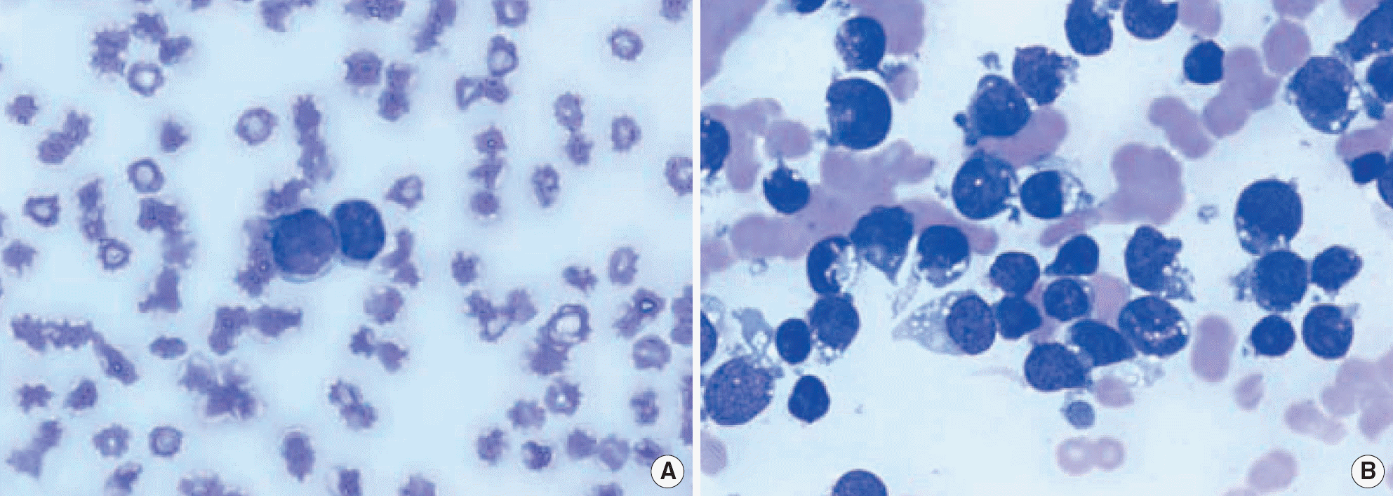
 | Fig. 1.Peripheral blood smear (A) showing blasts. Bone marrow smear (B) revealed many medium-sized lymphoblasts (about 82.4% of all nucleated cells) with moderate amount of cytoplasm, many vacuoles, and indistinct nucleoli (Wright-Giemsa stain, × 1,000). |
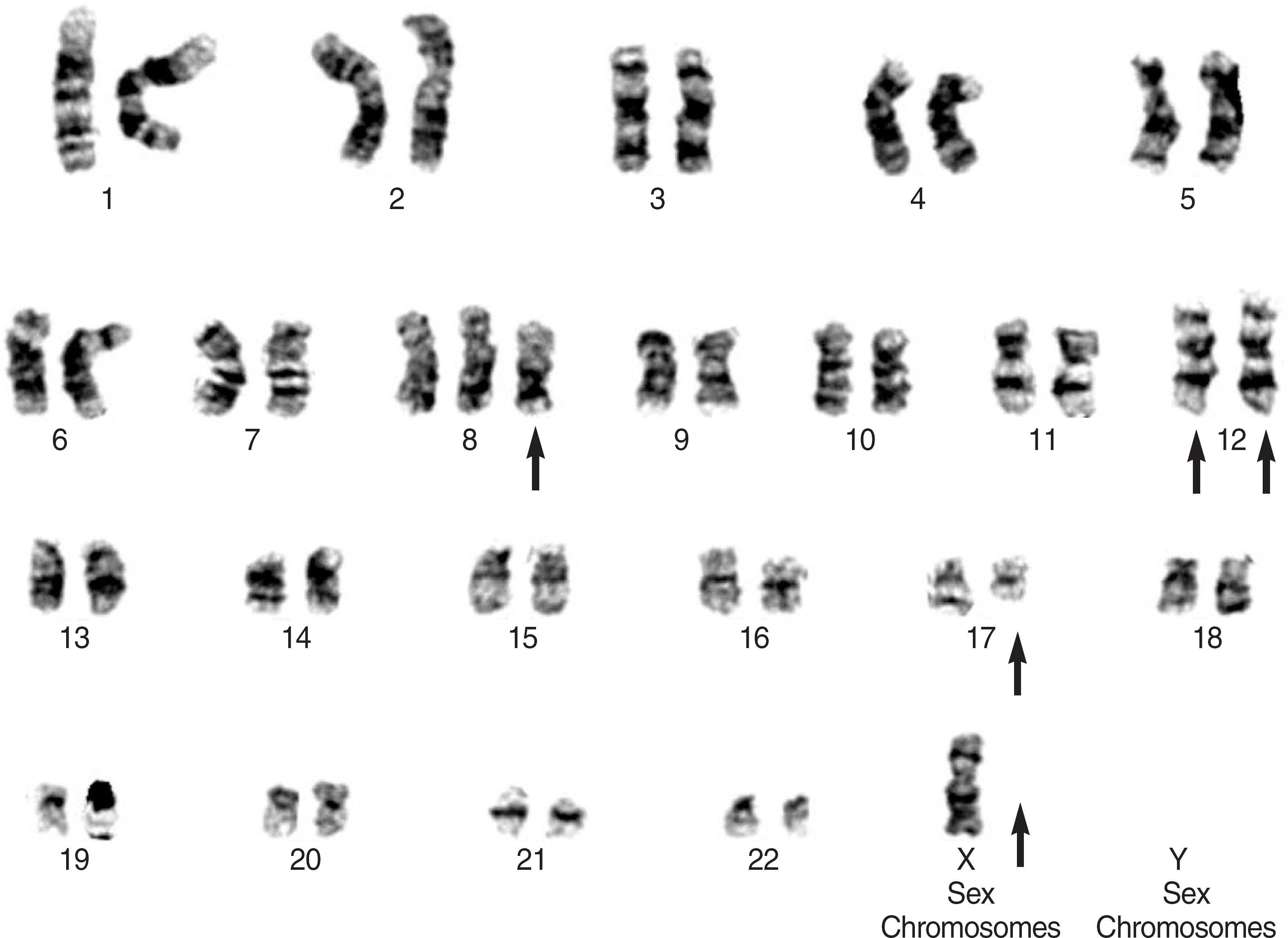
 | Fig. 2.Giemsa-banded karyotype showing 46,X,-X,+8,der(12)t(12;17)(p13;q21),t(12;17)(p13;q21) (arrows). |
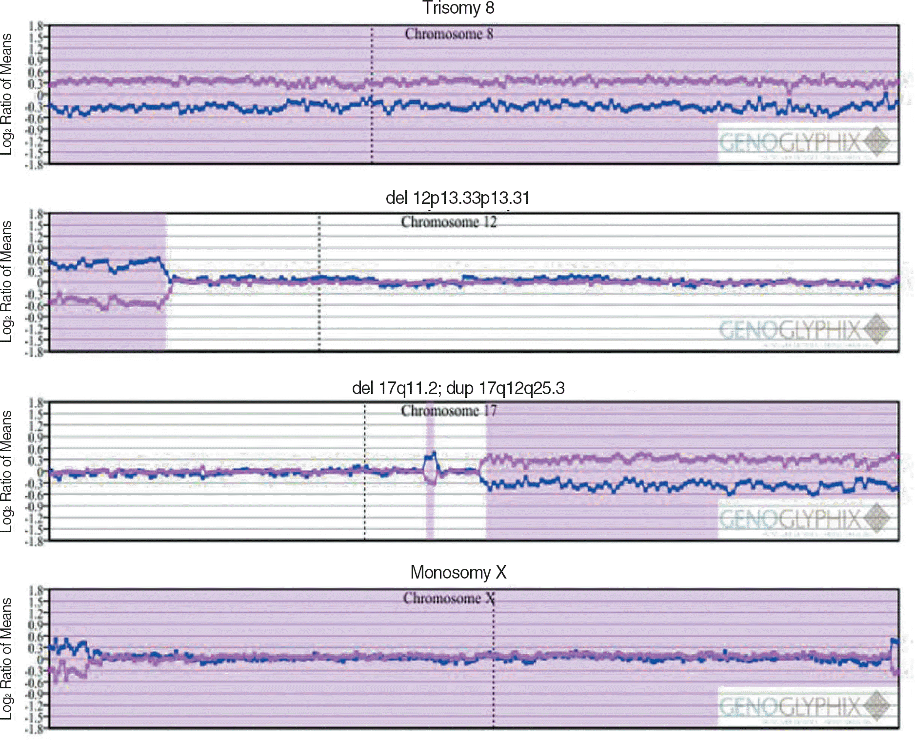
 | Fig. 3.The results of array comparative genomic hybridization were consistent with cytogenetic findings. The pink line represents the patient-to-control fluorescence intensity ratios, whereas the dark blue line represents dye-reversed control-to-patient fluorescence ratios. The microarray analysis revealed that the patient had 5 chromosomal abnormalities. The second abnormality is an approximately 6.6-Mb terminal deletion of the short arm of chromosome 12 at 12p13.33-12p13.31. The fourth abnormality is an approximately 47.4-Mb terminal duplication of the long arm of chromosome 17 at 17q12-17q25.3. |


 PDF
PDF ePub
ePub Citation
 Print
Print


 XML Download