Abstract
Tetraploidy or near-tetraploidy is a rare cytogenetic abnormality found in AML, and is divided into primary and secondary forms. The secondary tetraploidy or near-tetraploidy found in AML is known to be specifically associated with t(8;21). In this case report, FISH analysis detected RUNX1-RUNX1T1 gene rearrangement in the absence of cytogenetic abnormality of t(8;21), which suggests the presence of unvailed t(8;21). This is the first case report of tetraploidy or near-tetraploidy AML with cryptic RUNX1/RUNX1T1 in Korea. Although the prognosis of tetraploidy or near- tetraploidy with t(8;21) is known to be poor, this patient shows a relatively good clinical course compared to other reported cases.
Go to : 
REFERENCES
1.Secker-Walker LM., Prentice HG., Durrant J., Richards S., Hall E., Harrison G. Cytogenetics adds independent prognostic information in adults with acute lymphoblastic leukaemia on MRC trial UKALL XA. MRC adult leukaemia working party. Br J Haematol. 1997. 96:601–10.
2.Pui CH., Carroll AJ., Head D., Raimondi SC., Shuster JJ., Crist WM, et al. Near-triploid and near-tetraploid acute lymphoblastic leukemia of childhood. Blood. 1990. 76:590–6.

3.Mitelman F., Johansson B, et al. Mitelman database of chromosome aberrations in cancer. http://cgap.nci.nih.gov/Chromosomes/Mitelman. (Updated on May. 2009.
4.Iyer RV., Sait SN., Matsui S., Block AW., Barcos M., Slack JL, et al. Massive hyperdiploidy and tetraploidy in acute myelocytic leukemia and myelodysplastic syndrome. Cancer Genet Cytogenet. 2004. 148:29–34.

5.Xiao Z., Liu S., Liu X., Yu M., Hao Y. Tetraploidy or near-tetraploidy clones with double 8;21 translocation: a non-random additional anomaly of acute myeloid leukemia with t(8;21)(q22;q22). Haematologica. 2005. 90:413–4.
6.Testa JR., Oguma N., Pollak A., Wiernik PH. Near-tetraploid clones in acute leukemia. Blood. 1983. 61:71–8.

7.Abe R., Raza A., Preisler HD., Tebbi CK., Sandberg AA. Chromosomes and causation of human cancer and leukemia. LIV. Near-tetraploidy in acute leukemia. Cancer Genet Cytogenet. 1985. 14:45–59.

8.Xue Y., Pan Y., Liu Z., Li J., Guo Y., Xie X. Tetraploid or near-tetraploid clones characterized by two 8;21 translocations and other chromosomal abnormalities in two patients with acute myeloblastic leukemia. Cancer Genet Cytogenet. 1996. 92:18–23.
9.Xue Y., He J., Wang Y., Guo Y., Xie X., He Y, et al. Secondary near-pentaploidy and/or near-tetraploidy characterized by the duplication of 8;21 translocation in the M2 subtype of acute myeloid leukemia. Int J Hematol. 2000. 71:359–65.
10.Lemez P., Michalová K., Zemanová Z., Marinov I., Trpáková A., Moravcová J, et al. Three cases of near-tetraploid acute myeloid leukemias originating in pluripotent myeloid progenitors. Leuk Res. 1998. 22:581–8.
11.Yamamoto K., Nagata K., Kida A., Tsurukubo Y., Hamaguchi H. CD7+ near-tetraploid acute myeloblastic leukemia M2 with double t(8;21) (q22;q22) translocations and Aml1/ETO rearrangements detected by fluorescence in situ hybridization analysis. Int J Hematol. 2001. 74:316–21.
12.Udayakumar AM., Alkindi S., Pathare AV., Raeburn JA. Complex t(8;13;21)(q22;q14;q22)—a novel variant of t(8;21) in a patient with acute myeloid leukemia (AML-M2). Arch Med Res. 2008. 39:252–6.

13.Sarriera JE., Albitar M., Estrov Z., Gidel C., Aboul-Nasr R., Manshouri T, et al. Comparison of outcome in acute myelogenous leukemia patients with translocation (8;21) found by standard cytogenetic analysis and patients with AML1/ETO fusion transcript found only by PCR testing. Leukemia. 2001. 15:57–61.

14.Kim H., Kim M., Lim J., Kim Y., Han K., Kim SY, et al. A case of acute myeloid leukemia with masked t(8;21). Korean J Lab Med. 2006. 26:338–42. (김현정, 김명신, 임지향, 김용구, 한경자, 김성용등. Masked t(8;21)을 보이는 급성골수성백혈병 1예. 대한진단검사의학회지 2006;26:338-42.).

15.Swerdlow SH, Campo E, editors. WHO classification of tumours of haematopoietic and lymphoid tissues. 4th ed.Lyon: IARC Press;2008. p. 110–23.
16.Huret JL., Banerjee S, et al. Atlas of genetics and cytogenetics in oncology and haematology. http://atlasgeneticsoncology.org/index.html. (Updated on Jan. 2009.
Go to : 
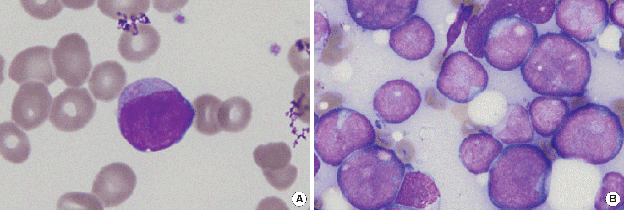
 | Fig. 1.Smear findings of the acute myeloid leukemia case. (A) Peripheral blood smear shows a large blast with Auer rods in the cytoplasm (Wright-Giemsa stain, ×1,000). (B) Bone marrow aspirate smear shows large blasts with irregular nuclear contours (Wright-Giemsa stain, ×1,000). |
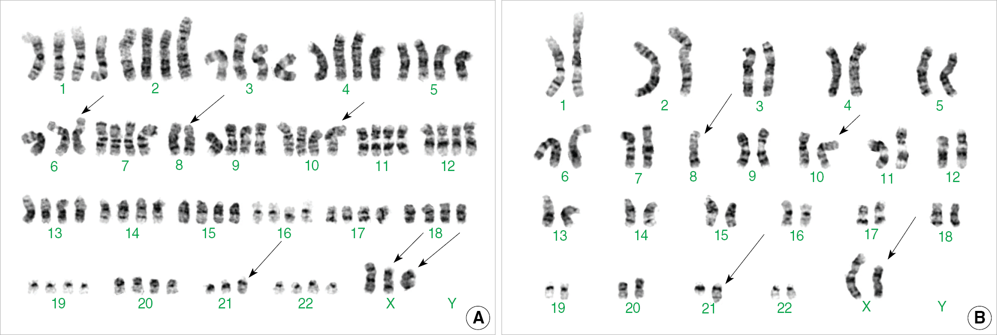
 | Fig. 2.Two representative karyotypes of the metaphases analyzed in the patient's bone marrow cells at the time of diagnosis. (A) shows 87,X,-X,add(X)(q13)×2,-6,-8,-8,add(10)(p13),-21,add(21)(q22) and (B) represents 45,X,-X,add(X)(q13),-8,add(10)(p13),add(21)(q22). |
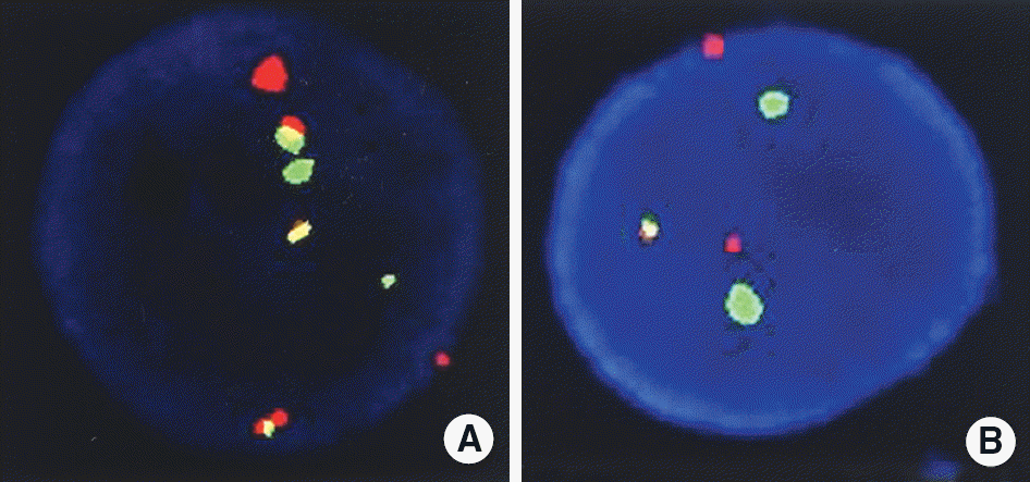
 | Fig. 3.
RUNX1-RUNX1T1 gene rearrangement detected by FISH. (A) Result of FISH analysis at the point of diagnosis. Shows an interphase cell with two orange (RUNX1T1) signals, two green (RUNX1) signals, and three fusion signals. (B) Result of FISH analysis two weeks after the diagnosis. An interphase cell has two orange (RUNX1T1) signals, two green (RUNX1) signals, and one fusion (RUNX1/ RUNX1T1) signal. |


 PDF
PDF ePub
ePub Citation
 Print
Print


 XML Download