Abstract
Background:
Near-infrared light (NIR, 0.8-1.5 μm light) has been used in therapeutic devices for various injuries such as infected, ischemic and hypoxic wound. NIR-emitting technology has been developed recently in Korea. We hypothesized that NIR may have an anti-inflammatory effect and investigated the effect of NIR-irradiated media on cell culture.
Methods:
Three kinds of cell lines, CAPE (vascular endothelial cell), NIH3T3 (fibroblast), and RD (smooth muscle cell) cells were cultured for 4 days in 10% FBS-containing media (1×10⁴ cells/well), which were irradiated or not irradiated (control) by Eco-NFIR Drive (Model #0210, Ecowavetech, Korea). The cells were stimulated by 10 mcg/mL of bacterial lipopolysaccharide (LPS) or sodium nitroprusside (SNP). Cellular proliferation was measured by methylthiazol tetrazolium assay. Expression of interleukin (IL)-1 beta and nitric oxide was measured by ELISA. Expression of cyclooxygenase-2 (COX-2) and inducible nitric oxide synthase (iNOS) was measured by immunofluorescence staining.
Results:
NIR-irradiated medium was favorable for CAPE cell proliferation (N=8, P=0.000). IL-1 beta secretion from LPS-stimulated NIH3T3 cells incubated in the NIR medium was below that of control medium (N=4, P=0.026). Nitrate production seemed to be low in NIR-irradiated medium although statistically insignificant (N=4, P=0.076). Expression of iNOS of the LPS-stimulated cells was decreased in NIR medium, however, Cox-2 expression was not different between the two media.
REFERENCES
1.Wong-Riley MT., Liang HL., Eells JT., Chance B., Henry MM., Buchmann E, et al. Photobiomodulation directly benefits primary neurons functionally inactivated by toxins: role of cytochrome c oxidase. J Biol Chem. 2005. 280:4761–71.
2.Eells JT., Wong-Riley MT., VerHoeve J., Henry M., Buchman EV., Kane MP, et al. Mitochondrial signal transduction in accelerated wound and retinal healing by near-infrared light therapy. Mitochondrion. 2004. 4:559–67.

3.Danno K., Mori N., Toda K., Kobayashi T., Utani A. Near-infrared irradiation stimulates cutaneous wound repair: laboratory experiments on possible mechanisms. Photodermatol Photoimmunol Photomed. 2001. 17:261–5.
4.Kandolf-Sekulovic L., Kataranovski M., Pavlovic MD. Immunomodulatory effects of low-intensity near-infrared laser irradiation on contact hypersensitivity reaction. Photodermatol Photoimmunol Photomed. 2003. 19:203–12.

5.Mochizuki-Oda N., Kataoka Y., Cui Y., Yamada H., Heya M., Awazu K. Effects of near-infra-red laser irradiation on adenosine triphosphate and adenosine diphosphate contents of rat brain tissue. Neurosci Lett. 2002. 323:207–10.

6.Oh WS., In JY., Ha KH., Hong KH. Application of polarized light irradiation to meralgia paresthetica. Korean J Anesthesiol. 2000. 38:183–6. (오완수, 인준용, 하경호, 홍기혁. 직선편광 근적외선 조사에 의한 지각이상성대퇴신경통의치료경험. 대한마취과학회지 2000;38:183-6.).

7.Han SM., Lee SC. The change of blood flow velocity of radial artery after linear polarized infrared light radiation near the stellate ganglion: comparing with the stellate ganglion block. Korean J Pain. 2001. 14:37–40. (한승문 및 이상철. 성상신경절 부위의 직선편광 근적외선조사후요골동맥에서의혈류속도의변화: 성상신경절차단술과의비
교. 대한통증학회지 2001;14:37-40.).
8.Kim J., Otzel D., Kim W., Janelle CM. Near-infrared light and expectancy effects on maximal isokinetic strength performance: a randomized, double-blind, placebo-controlled study. J Strength Cond Res. 2006. 20:378–82.

9.Choi CH., Lee HS., Seo HS., Kim SG., Shin IH. Biochemical changes and recovery after half-course marathon. J Korean Orthop Soc Sports Med. 2008. 7:45–9. (최창혁, 이현섭, 서헌석, 김상경, 신임희. 하프코스마라톤후체내의생화학적변화및회복. 대한정형외과스포츠의학회지 2008;7:45-9.).
10.Kim SS., Kim WJ., Kim HE. The study of thermo-physiological responses with near infrared lighted garment at a hot environment. J Korean Soc Cloth Ind. 2005. 7:665–72. (김성숙, 김우종, 김희은. 서열환경에서 근적외선 조사의복 착용시의 온열생리반응. 한국의류산업학회지 2005; 7:665–72.).
11.Park KH., Park YM., Seul KJ., Ghim SY. The indirect effects of the near infra-red light-treated materials on microbial growth. Korean J Microbiol Biotechnol. 2005. 33:222–5. (박경화, 박유미, 설경조, 김사열. 근적외선을 처리한 생활용품의 항균 효과. 한국미생물생명공학회지 2005;33:222-5.).
12.Song JS. Review of tumor necrosis factor inhibitors on rheumatoid arthritis. J Korean Rheum Assoc. 2007. 14:1–14. (송정수. 류마티스관절염 치료에서 종양괴사인자억제제 사용에 대한 고찰. 대한류마티스학회지 2007;14:1-14.).

13.Denizot F., Lang R. Rapid colorimetric assay for cell growth and survival. Modifications to the tetrazolium dye procedure giving improved sensitivity and reliability. J Immunol Method. 1986. 89:271–7.
14.Kim SG., Choe JY., Choi CH., Choi GH., Kim JK. Effect of culture condition on chondrocyte viability isolated from articular cartilage. Korean J Lab Med. 2004. 24:237–43. ((김상경, 최정윤, 최창혁, 최기환, 김종기. 관절연골조직으로부터 연골세포 일차배양시 배양방법에 따른 세포의생존력. 대한진단검사의학회지 2004;24:237-43.).
15.Kim S., Temple MM., Chen AC., Sah RL. Effect of nitric oxide on cartilage homeostasis and chondrocyte viability. J Orhto Res. 2001. ;S18.
16.Komorowska M., Cuissot A., Czarnoleski A., Bialas W. Erythrocyte response to near-infrared radiation. J Photochem Photobiol B. 2002. 68:93–100.

17.Whelan HT., Connelly JF., Hodgson BD., Barbeau L., Post AC., Bullard G, et al. NASA light-emitting diodes for the prevention of oral mucositis in pediatric bone marrow transplant patients. J Clin Laser Med Surg. 2002. 20:319–24.

18.Whelan HT., Smits RL Jr., Buchman EV., Whelan NT., Turner SG., Margolis DA, et al. Effect of NASA light-emitting diodes irradiation on wound healing. J Clin Laser Med Surg. 2001. 19:305–14.
Fig. 1.
Methylthiazol tetrazolium test result of the cells. NIR-irradiated medium provided favorable condition for CAPE cells in terms of cellular mitochondrial enzyme (succinate dehydrogenase) actvity (mean±SE, N=8, P=0.000). However, there was no significant difference of cellular proliferation of NIH3T3 cell (mean±SE, N=16, P=0.170) and RD cell (mean±SE, N=8, P=0.295) between NIR-irradiated medium (N, –Δ–) and control medium (C, –○–).
Abbreviations: C, control medium; N, NIR-irradiated medium; MTT, methylthiazol tetrazolium; NIR, near-infrared irradiated.

Fig. 2.
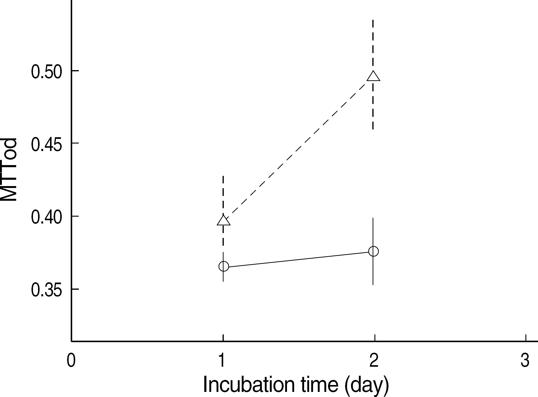
Methylthiazol tetrazolium test result of SNP-stimulated NIH3T3 cells. The SNP-stimulated NIH3T3 cells incubated in the NIR-irradiated medium (–Δ–) proliferated more than the cells incubated in the control medium (–○–) at day 2 (mean±SE, N=16, P=0.000). Abbreviations: See Fig. 1.

Fig. 3.
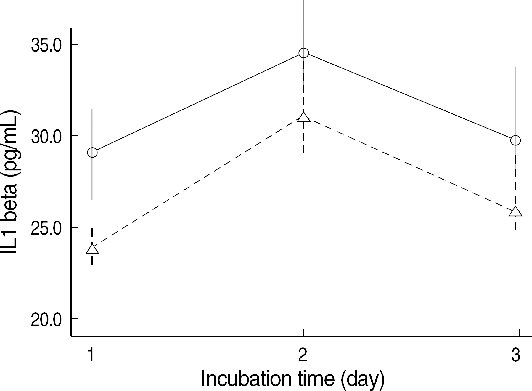
IL-1 beta concentration of the supernatant of NIH3T3 cells which were stimulated with LPS (10 mcg/mL). The IL-1 beta secretion of NIH3T3 cells incubated in NIR-irradiated medium (–Δ–) was below that in the control medium (–○–) (mean±SE, N=4, P=0.026).
Abbreviations: See Fig. 1.

Fig. 4.
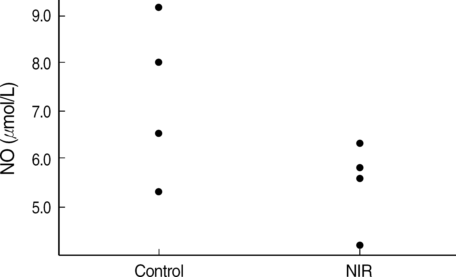
Nitric oxide production of NIH3T3 cells which were stimulated with LPS (10 mcg/mL). The NO level of NIH3T3 cells incubated in NIR-irradiated medium (NIR) was lower than that in the control medium (control), although statistically insignificant (mean±SE, N=4, P=0.076).
Abbreviations: See Fig. 1.

Fig. 5.
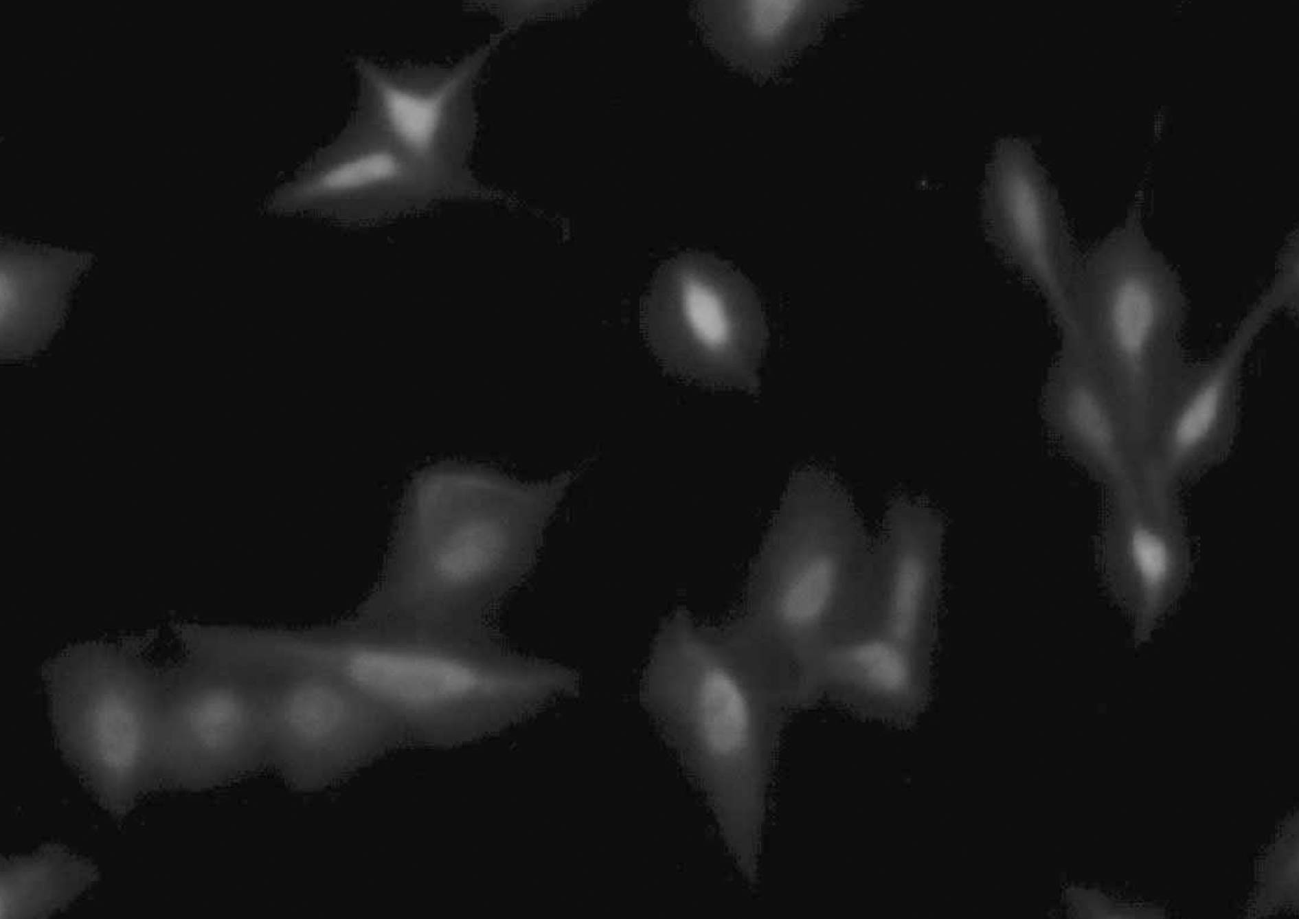
LPS (10 mcg/mL) stimulated NIH3T3 cells show green fluorescence after Immunofluorescence staining of iNOS-FITC antibody (epi-fluoresence microscopy, NiKon, micro-FXA ×400). Abbreviations: See Table 1.

Table 1.
Result of immunofluorescence staining of inducible nitric oxide synthase expression of the NIH3T3 cells


 PDF
PDF ePub
ePub Citation
 Print
Print


 XML Download