

 PDF
PDF ePub
ePub Citation
 Print
Print


Abstract
Histiocytic sarcoma is a malignant proliferation of cells showing morphologic and immunophenotypic features similar to those of mature tissue histiocytes and is known for its rapid progression and poor prognosis. We describe a case of histiocytic sarcoma diagnosed by bone marrow biopsy. A 64-yr-old male was admitted for fever and weight loss that persisted for 8 months. The patient died undiagnosed on the 7th hospitalization day. A bone marrow biopsy performed just before the patient's death revealed diffuse proliferation of large pleomorphic neoplastic cells with large, round to oval nuclei, vesicular chromatin, and abundant foamy cytoplasm. These cells were positive for histiocytic markers, CD68, lysozyme, CD21, and S-100 protein, but negative for B-cell, T/NK-cell, and epithelial cell markers, thus confirming the presence of histiocytic sarcoma.
REFERENCES
1.Jaffe ES., Harris NL, et alWorld Health Organization Classification of tumours: pathology and genetics of tumours of haematopoietic and lymphoid tissues. Lyon: IARC Press. 2008. 356–7.
2.Yoshida C., Takeuchi M. Histiocytic sarcoma: identification of its histiocytic origin using immunohistochemistry. Intern Med. 2008. 47:165–9.

3.Pileri SA., Grogan TM., Harris NL., Banks P., Campo E., Chan JK, et al. Tumours of histiocytes and accessory dendritic cells: an immuno-histochemical approach to classification from the International Lymphoma Study Group based on 61 cases. Histopathology. 2002. 41:1–29.
4.Vos JA., Abbondanzo SL., Barekman CL., Andriko JW., Miettinen M., Aguilera NS. Histiocytic sarcoma: a study of five cases including the histiocyte marker CD163. Mod Pathol. 2005. 18:693–704.
5.Soria C., Orradre JL., Garcia-Almagro D., Martinez B., Algara P., Piris MA. True histiocytic lymphoma (monocytic sarcoma). Am J Dermatopathol. 1992. 14:511–7.
6.deMent SH. Association between mediastinal germ cell tumors and hematologic malignancies: an update. Hum Pathol. 1990. 21:699–703.
7.Park MI., Song KS., Kang DY. Histiocytic sarcoma of rectum: a case report. Korean J Pathol. 2006. 40:156–9.
8.Paik JH., Jeon YK., Park SS., Min HS., Kim YA., Kim JE, et al. Histiocytic sarcoma of the spleen: a case report and review of the literature. Korean J Pathol. 2005. 39:356–9.
9.Kim KW., Park SY., Kim HJ., Yoo BH., Kang SB., Lee JP, et al. A case with disseminated macrophage-related histiocytic sarcoma diagnosed by positive histiocytic markers. Korean J Hematol. 1999. 34:641–5. (김기원, 박석영, 김희정, 유병현, 강상범, 이재필 등. 조직구 표지자 염색양성으로 확인된범발성 대식세포관련조직구육종1예. 대한혈액학회지 1999;34: 641-5.).
10.Hornick JL., Jaffe ES., Fletcher CD. Extranodal histiocytic sarcoma: clinicopathologic analysis of 14 cases of a rare epitheloid malignancy. Am J Surg Pathol. 2004. 28:1133–44.
Fig. 1.
Morphologic and immunohistochemical features of the peripheral blood and bone marrow specimen. (A) Rouleaux formation in the peripheral blood smear (Wright stain, ×1,000). (B) Biopsy specimen showing high cellularity, diffuse fibrosis, and large pleomorphic neoplastic cells (H&E stain, ×400). (C) Biopsy specimen showing CD31+ (immunohistochemistry, ×400). (D) Biopsy specimen showing CD68+ cells (immunohistochemistry, ×400). (E) Biopsy specimen showing lysozyme-positive cells (immunohistochemistry, ×400). (F) Biopsy specimen showing S100 protein-positive cells (immunohistochemistry, ×400).
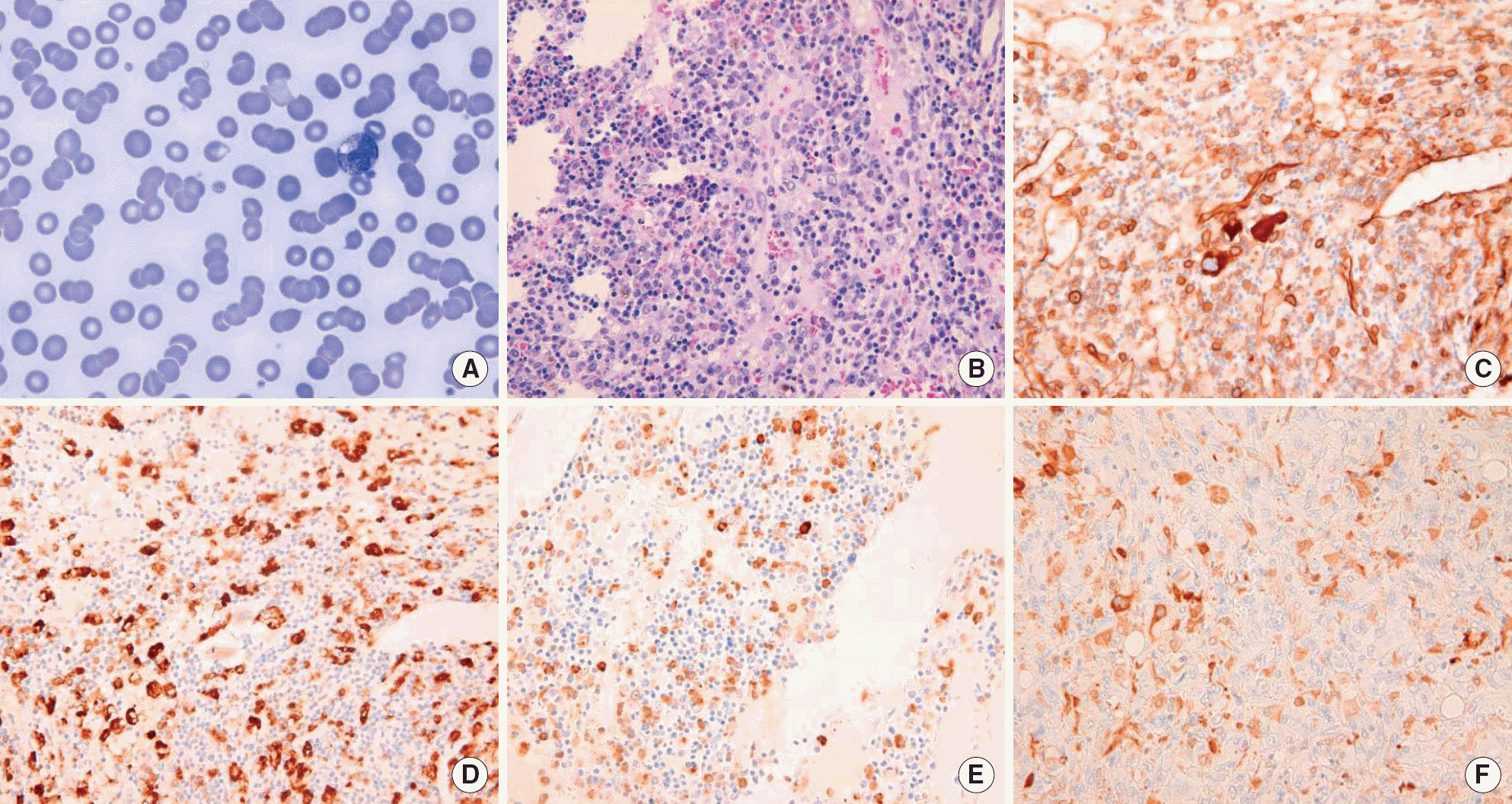
Table 1.
Comparison of the immunohistochemical features of histiocytic sarcoma in this case and several other reported cases
 XML Download