Abstract
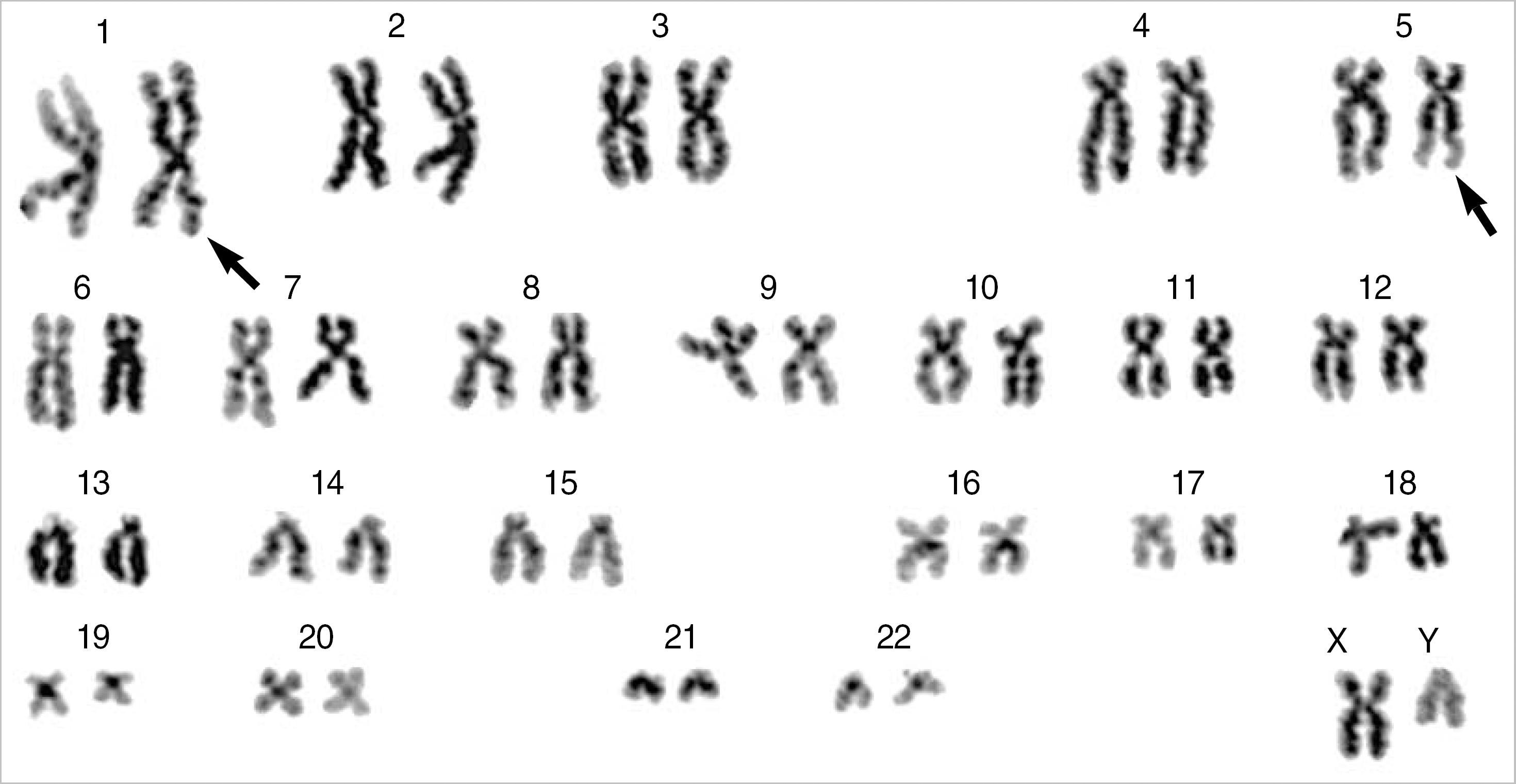
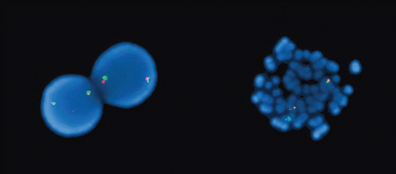
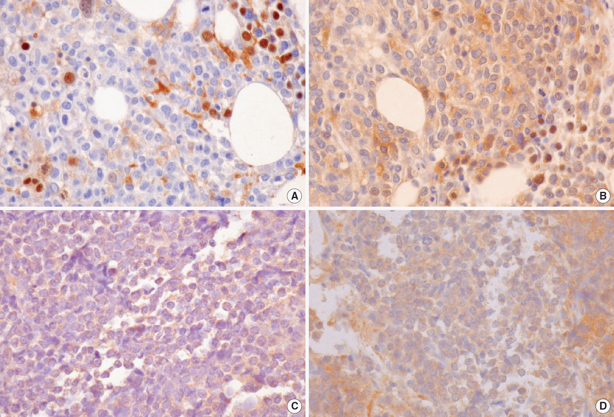
Chromosome 1 band p32 (1p32) aberrations are common in T lymphoblastic leukemia/lymphoma (T-ALL/LBL). Two types of 1p32 aberrations include translocations with different partners and sub-microscopic interstitial deletion. Both aberrations are known to result in TAL1 gene deregulation. The t(1;5)(p32;q31) is a rare translocation of 1p32 in T-ALL. We now present the second case of t(1;5)(p32;q31) in T-ALL, which was present as a primary cytogenetic abnormality, with a review of the relevant literature. Interestingly, neither the translocation of the TAL1 gene nor aberrant expression of TAL1 protein was detected by fluorescent in situ hybridization (FISH) and by immunohistochemical staining in this case.
REFERENCES
1.Swerdlow SH, Campo E, editors. WHO classification of tumours of haematopoietic and lymphoid tissues. 4th ed.Lyon: IARC press;2008. p. 14–5.
2.Mitelman F, Johansson B, Mertens F, editors. Mitelman database of chromosome aberrations in cancer. http://cgap.nci.nih.gov/Chromosomes/Mitelman. (Updated on Nov. 2008.
3.Carroll AJ., Crist WM., Link MP., Amylon MD., Pullen DJ., Ragab AH, et al. The t(1;14)(p34;q11) is nonrandom and restricted to T-cell acute lymphoblastic leukemia: a Pediatric Oncology Group study. Blood. 1990. 76:1220–4.

4.Fitzgerald TJ., Neale GA., Raimondi SC., Goorha RM. c-tal, a helix-loop-helix protein, is juxtaposed to the T-cell receptor β chain gene by a reciprocal chromosomal translocation: t(1;7)(p32;q35). Blood. 1991. 78:2686–95.
5.Aplan PD., Raimondi SC., Kirsch IR. Disruption of the SCL gene by a t(1;3) translocation in a patient with T cell acute lymphoblastic leukemia. J Exp Med. 1992. 176:1303–10.

6.François S., Delabesse E., Baranger L., Dautel M., Foussard C., Boasson M, et al. Deregulated expression of the TAL1 gene by t(1;5)(p32;q31) in patient with T-cell acute lymphoblastic leukemia. Genes Chromosomes Cancer. 1998. 23:36–43.
7.Kaleem Z., Shuster JJ., Carroll AJ., Borowitz MJ., Pullen DJ., Camitta BM, et al. Acute lymphoblastic leukemia with an unusual t(8;14)(q11.2;q32): a Pediatric Oncology Group study. Leukemia. 2000. 14:238–40.

8.van der Burg M., Poulsen TS., Hunger SP., Beverloo HB., Smit EM., Vang-Nielsen K, et al. Split-signal FISH for detection of chromosome aberrations in acute lymphoblastic leukemia. Leukemia. 2004. 18:895–908.

9.Pulford K., Lecointe N., Leroy-Viard K., Jones M., Mathieu-Mahul D., Mason DY. Expression of TAL-1 proteins in human tissues. Blood. 1995. 85:675–84.

10.Finger LR., Kagan J., Christopher G., Kurtzberg J., Hershfield MS., Nowell PC, et al. Involvement of the TCL5 gene on human chromosome 1 in T-cell leukemia and melanoma. Proc Natl Acad Sci USA. 1989. 86:5039–43.

11.Brown L., Cheng JT., Chen Q., Siciliano MJ., Crist W., Buchanan G, et al. Site-specific recombination of the tal-1 gene is a common occurrence in human T cell leukemia. EMBO J. 1990. 9:3343–51.

12.Huret JL. Atlas of genetics and cytogenetics in oncology and haematology. http://atlasgeneticsoncology.org. (Updated on Oct. 2008.
13.Bash RO., Crist WM., Shuster JJ., Link MP., Amylon M., Pullen J, et al. Clinical features and outcome of T-cell acute lumphoblastic leukemia in childhood with respect to alterations at the TAL1 locus: a Pediatric Oncology Group study. Blood. 1993. 81:2110–7.
14.van Grotel M., Meijerink JP., van Wering ER., Langerak AW., Beverloo HB., Buijs-Gladdines JG, et al. Prognostic significance of molecular-cytogenetic abnormalities in pediatric T-ALL is not explained by immunophenotypic differences. Leukemia. 2008. 22:124–31.

15.Cotterill S. Cancer Genetics Web. http://www.cancer-genetics.org. (Updated on Apr. 2003.
Fig. 1.
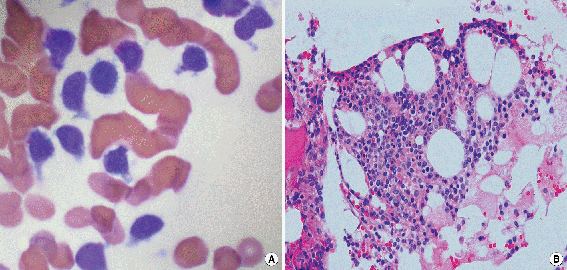
(A) The bone marrow aspirates (Wright stain, ×1,000) and (B) biopsy (hematoxylin and eosin stain, ×200) showed an increase of small to medium sized blasts.


 PDF
PDF ePub
ePub Citation
 Print
Print


 XML Download