Abstract
Background
Cardiac troponin I (cTnI) is known as a sensitive and specific marker for myocardial ischemia. The purposes of this study are to establish cut-off values of cTnI for acute myocardial infarction (AMI) and to analyze clinical significance of minor elevation of cTnI.
Methods
Two hundred and four patients from whom cTnI was measured at Ewha Womans University Dongdaemun hospital from January to March, 2006 were enrolled in the study. cTnI was measured using Dimension RxL (Dade Behring, USA). The lower limit of detection (LLD), 10% CV value, 99th percentile of healthy individuals, and cut-off value for AMI by ROC curve analysis were determined.
Results
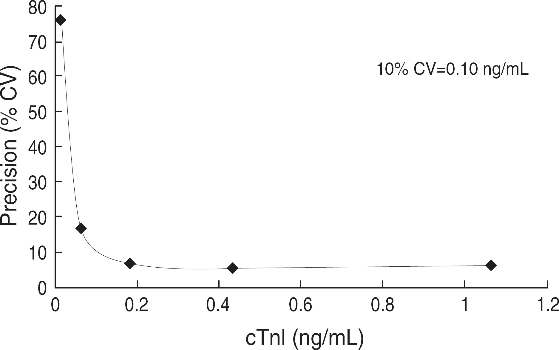
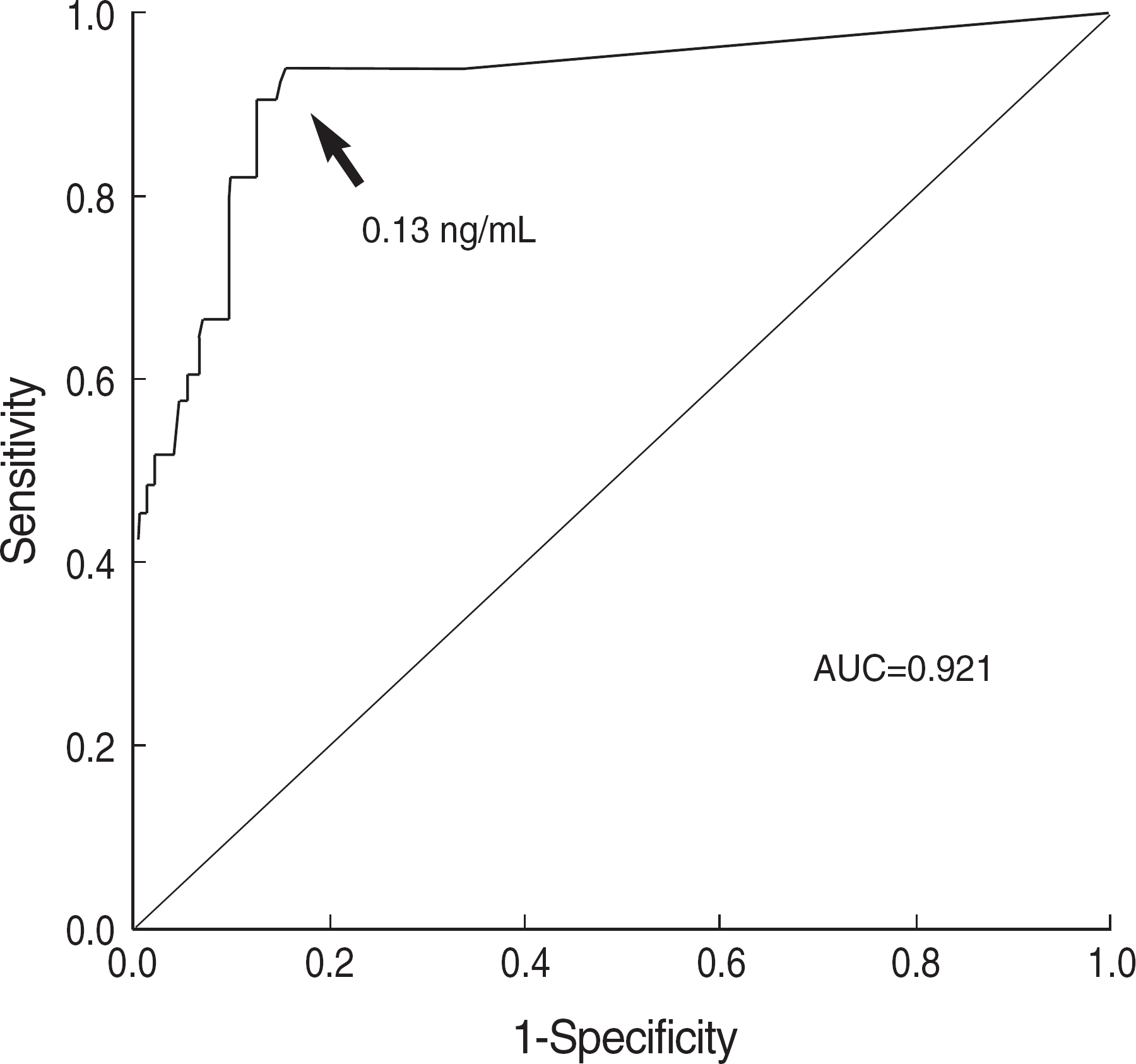
LLD, 10% CV value, and 99th percentile of cTnI were 0.00 ng/mL, 0.10 ng/mL, and 0.07 ng/mL, respectively. The cut-off value of peak cTnI for AMI by ROC curve analysis was 0.13 ng/mL with the sensitivity, specificity, and AUC of 90.9%, 87.7%, and 0.921, respectively. The peak value of cTnI of patients with ischemic heart disease (IHD) was higher than that of the patients without IHD (P<0.05). According to the above reference and cut-off values of the initial cTnI, patients were categorized into four groups; ≤0.05 ng/mL (group 1), 0.06-0.09 ng/mL (group 2), 0.10-0.59 ng/mL (group 3), ≥0.60 ng/mL (group 4), and compared frequencies of AMI, IHD, cardio vascular disease (CVD) and death after 1 month among groups. Frequencies of AMI, IHD, CVD, and death after 1 month were significantly increased as the cTnI concentrations were increased (P<0.05).
Go to : 
REFERENCES
1.Myocardial infarction redefined–a consensus document of The Joint European Society of Cardiology/American College of Cardiology Committee for the redefinition of myocardial infarction. Eur Heart J. 2000. 21:1502–13.
2.Jaffe AS., Ravkilde J., Roberts R., Naslund U., Apple FS., Galvani M, et al. It's time for a change to a troponin standard. Circulation. 2000. 102:1216–20.

3.Babuin L., Jaffe AS. Troponin: the biomarker of choice for the detection of cardiac injury. CMAJ. 2005. 173:1191–202.

4.Gudmundsson GS., Kahn SE., Moran JF. Association of mild transient elevation of troponin I levels with increased mortality and major cardiovascular events in the general patient population. Arch Pathol Lab Med. 2005. 129:474–80.

5.Apple FS., Wu AH. Myocardial infarction redefined: role of cardiac troponin testing. Clin Chem. 2001. 47:377–9.

6.Morrow DA., Cannon CP., Rifai N., Frey MJ., Vicari R., Lakkis N, et al. Ability of minor elevations of troponins I and T to predict benefit from an early invasive strategy in patients with unstable angina and non-ST elevation myocardial infarction: results from a randomized trial. JAMA. 2001. 286:2405–12.
7.Braunwald E., Antman EM., Beasley JW., Califf RM., Cheitlin MD., Hochman JS, et al. ACC/AHA guideline update for the management of patients with unstable angina and non-ST-segment elevation myocardial infarction–2002: summary article: a report of the American College of Cardiology/American Heart Association Task Force on Practice Guidelines (Committee on the Management of Patients With Unstable Angina). Circulation. 2002. 106:1893–900.
8.Zarich SW., Bradley K., Mayall ID., Bernstein LH. Minor elevation in troponin T values enhance risk assessment in emergency department patients with suspected myocardial ischemia: analysis of novel troponin T cut-off values. Clin Chim Acta. 2004. 343:223–9.
9.Nomenclature and criteria for diagnosis of ischemic heart disease. Report of the Joint International Society and Federation of Cardiology/World Health Organization task force on standardization of clinical nomenclature. Circulation. 1979. 59:607–9.
10.Apple FS., Wu AH., Jaffe AS. European Society of Cardiology and American College of Cardiology guidelines for redefinition of myocardial infarction: how to use existing assays clinically and for clinical trials. Am Heart J. 2002. 144:981–6.

11.Panteghini M. Present issues in the determination of troponins and other markers of cardiac damage. Clin Biochem. 2000. 33:161–6.

12.Clinical and Laboratory Standards Institute. User demonstration of performance for precision and accuracy; approved guideline. Document EP15-A. Wayne, PA: Clinical and Laboratory Standards Institute;2001.
13.Ferguson JL., Beckett GJ., Stoddart M., Walker SW., Fox KA. Myocardial infarction redefined: the new ACC/ESC definition, based on cardiac troponin, increases the apparent incidence of infarction. Heart. 2002. 88:343–7.

14.Polanczyk CA., Schneid S., Imhof BV., Furtado M., Pithan C., Rohde LE, et al. Impact of redefining acute myocardial infarction on incidence, management and reimbursement rate of acute coronary syndromes. Int J Cardiol. 2006. 107:180–7.

15.Kavsak PA., MacRae AR., Lustig V., Bhargava R., Vandersluis R., Palomaki GE, et al. The impact of the ESC/ACC redefition of myocardial infarction and new sensitive troponin assays on the frequency of acute myocardial infarction. Am Heart J. 2006. 152:118–25.
16.Panteghini M., Pagani F., Yeo KT., Apple FS., Christenson RH., Dati F, et al. Evaluation of imprecision for cardiac troponin assays at low-range concentrations. Clin Chem. 2004. 50:327–32.

17.Melanson SE., Morrow DA., Jarolim P. Earlier detection of myocardial injury in a preliminary evaluation using a new troponin I assay with improved sensitivity. Am J Clin Pathol. 2007. 128:282–6.

18.Makaryus AN., Makaryus MN., Hassid B. Falsely elevated cardiac troponin I levels. Clin Cardiol. 2007. 30:92–4.

19.Kontos MC., Shah R., Fritz LM., Anderson FP., Tatum JL., Ornato JP, et al. Implication of different cardiac troponin I levels for clinical outcomes and prognosis of acute chest pain patients. J Am Coll Cardiol. 2004. 43:958–65.

20.Lindahl B., Diderholm E., Lagerqvist B., Venge P., Wallentin L. Mechanisms behind the prognostic value of troponin T in unstable coronary artery disease: a FRISC II substudy. J Am Coll Cardiol. 2001. 38:979–86.

21.Henrikson CA., Howell EE., Bush DE., Miles JS., Meininger GR., Friedlander T, et al. Prognostic usefulness of marginal troponin T elevation. Am J Cardiol. 2004. 93:275–9.

22.Cook G., Taylor D., France M., Burrows G., Manning E., Lyratzopoulos G, et al. Survival among hospital in-patients with troponin T elevation below levels defining myocardial infarction. QJM. 2005. 98:275–82.

23.Lindahl B., Venge P., Wallentin L. Troponin T identifies patients with unstable coronary artery disease who benefit from long-term anti-thrombotic protection. Fragmin in Unstable Coronary Artery Disease (FRISC) Study Group. J Am Coll Cardiol. 1997. 29:43–8.
Go to : 
 | Fig. 1.Total precision profile for cardiac troponin I (cTnI). The 10% CV value was 0.10 ng/mL. |
 | Fig. 2.ROC curve for cTnI in 204 patients with or without AMI. Abbreviations: AUC, area under the curve; cTnI, cardiac troponin I; AMI, acute myocardial infarction. |
Table 1.
The number of AMI by cTnI cut-off of 0.13 ng/mL
| cTnI | AMI (N=33) | No AMI (N=171) |
|---|---|---|
| ≥0.13 ng/mL | 30 | 21 |
| <0.13 ng/mL | 3 | 150 |
Table 2.
Comparison of laboratory parameters between no IHD population and IHD population
| No IHD (N=140) Mean±SD | IHD (N=64) Mean±SD | P value∗ | |
|---|---|---|---|
| Age (yr) | 64.0±14.4 | 66.8±13.5 | 0.181 |
| cTnI (ng/mL) | |||
| Initial value | 0.06±0.06 | 1.67±8.27 | 0.123 |
| Peak value† | 0.09±0.20 | 4.94±15.02 | 0.012 |
| CK (U/L) | 491.3±2846.1 | 395.6±952.8 | 0.793 |
| CK-MB (ng/mL) | 19.3±145.5 | 10.7±35.6 | 0.641 |
| LD (U/L) | 533.5±305.6 | 575.7±214.3 | 0.335 |
| BNP (pg/mL) | 237.5±662.7 | 538.8±975.4 | 0.077 |
| Glucose (mg/dL) | 140.0±61.9 | 175.7±142.1 | 0.062 |
| BUN (mg/dL)† | 19.0±11.4 | 23.7±12.6 | 0.008 |
| Cr (mg/dL) | 1.34±4.50 | 1.34±1.63 | 0.999 |
| CRP (mg/dL) | 5.6±9.4 | 4.9±6.1 | 0.676 |
| TG (mg/dL) | 112.8±61.0 | 106.6±59.3 | 0.572 |
| LDL-C (mg/dL) | 114.2±47.4 | 101.2±46.3 | 0.241 |
| HDL-C (mg/dL)† | 48.1±19.9 | 39.5±12.2 | 0.030 |
| Total cholesterol (mg/dL) | 178.5±49.7 | 166.1±45.3 | 0.156 |
Abbreviations: IHD, ischemic heart disease; cTnI, cardiac troponin I CK, creatine kinase; LD, lactate dehydrogenase; BNP, B-type natriuretic peptide; CRP, C-reactive protein; BUN, blood urea nitrogen; Cr, creatinine; TG, triglyceride; LDL-C, low density lipoprotein-cholesterol; HDL-C, high density lipoprotein-cholesterol
Table 3.
Incidence of AMI, IHD, CVD, and death according to initial cTnI values
| cTnI (ng/mL) | P value∗ | ||||
|---|---|---|---|---|---|
| Group 1 ≤0.05 (N=142) [%, (N)] | Group 2 0.06-0.09 (N=22) [%, (N)] | Group 3 0.10-0.59 (N=30) [%, (N)] | Group 4 ≥0.60 (N=10) [%, (N)] | ||
| No AMI (N=171) | 95.8 (136) | 81.8 (18) | 46.7 (14) | 30.0 (3) | ≤0.001 |
| AMI (N=33) | 4.2 (6) | 18.2 (4) | 53.3 (16) | 70.0 (7) | |
| No IHD (N=140) | 81.0 (115) | 68.2 (15) | 30.0 (9) | 10.0 (1) | ≤0.001 |
| IHD (N=64) | 19.0 (27) | 31.8 (7) | 70.0 (21) | 90.0 (9) | |
| No CVD (N=116) | 70.4 (100) | 50.0 (11) | 13.3 (4) | 10.0 (1) | ≤0.001 |
| CVD (N=88) | 29.6 (42) | 50.0 (11) | 86.7 (26) | 90.0 (9) | |
| Survival (N=134) | 93.7 (104) | 78.6 (11) | 63.2 (12) | 70.0 (7) | ≤0.001 |
| Death (N=20) | 6.3 (7) | 21.4 (3) | 36.8 (7) | 30.0 (3) | |
Table 4.
Results of laboratory parameters according to initial cTnI values
| cTnI (ng/mL) | P value∗ | ||||
|---|---|---|---|---|---|
| Group 1 ≤0.05 (N=142) [%, (N)] | Group 2 0.06-0.09 (N=22) [%, (N)] | Group 3 0.10-0.59 (N=30) [%, (N)] | Group 4 ≥0.60 (N=10) [%, (N)] | ||
| CK (U/L) | 169.6±474.6 | 194.2±251.3 | 1,974.2±6008.2 | 587.8±782.8 | 0.002 |
| CK-MB (ng/mL) | 1.7±6.6 | 3.4±3.9 | 86.1±308.9 | 48.3±81.5 | ≤0.001 |
| LD (U/L) | 504.8±247.1 | 548.3±249.8 | 696.8±393.0 | 648.5±148.4 | ≤0.001 |
| BNP (pg/mL) | 176.2±434.5 | 117.5±126.6 | 816.3±1,266.7 | 1,048.5±1,450.3 | ≤0.001 |
| BUN (pg/mL) | 19.0±10.7 | 20.2±17.0 | 24.8±12.3 | 27.9±9.7 | 0.003 |
| Cr (mg/dL) | 1.00±0.93 | 3.22±11.1 | 1.40±1.70 | 1.82±1.61 | 0.007 |


 PDF
PDF ePub
ePub Citation
 Print
Print


 XML Download