Abstract
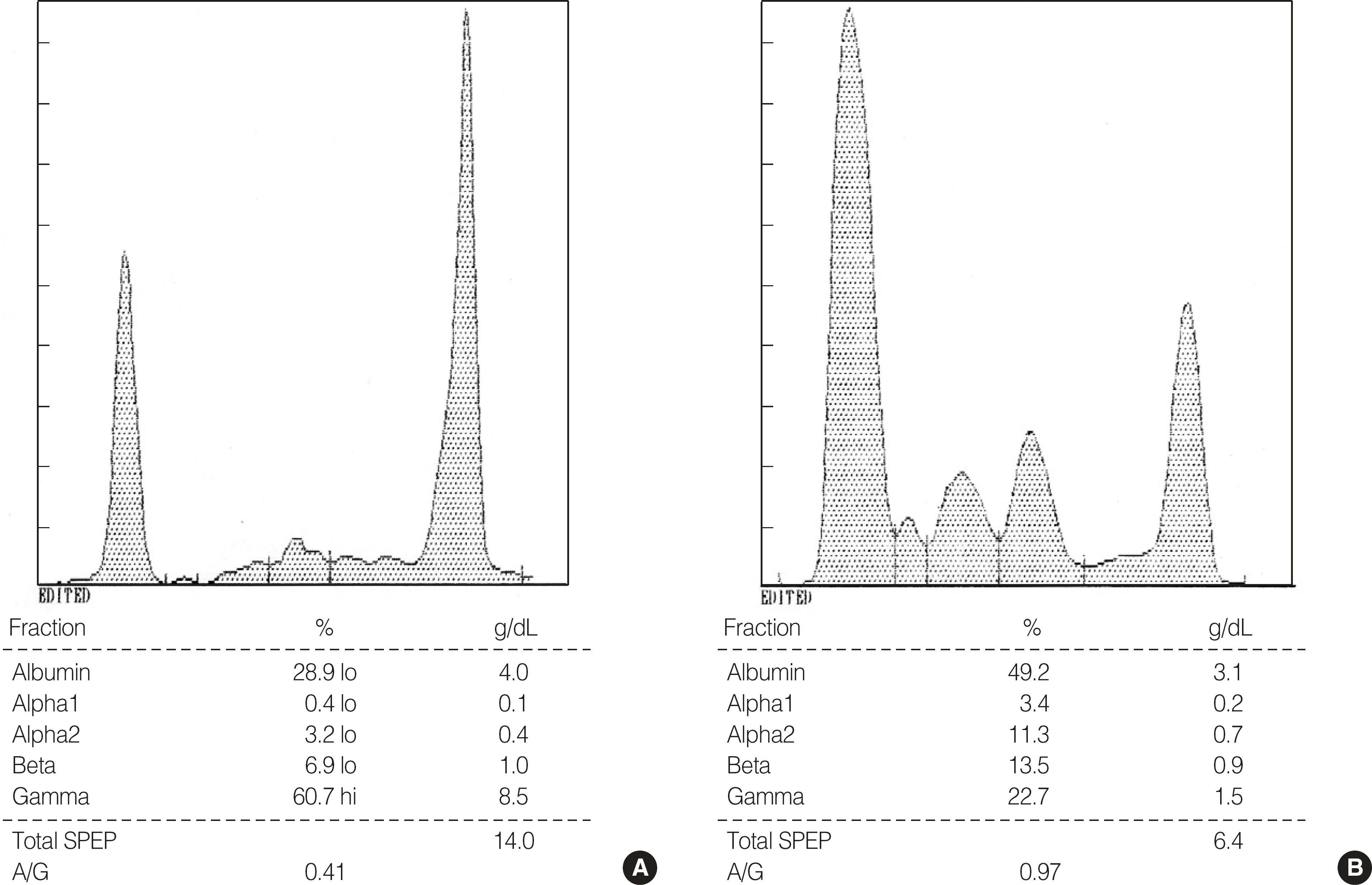
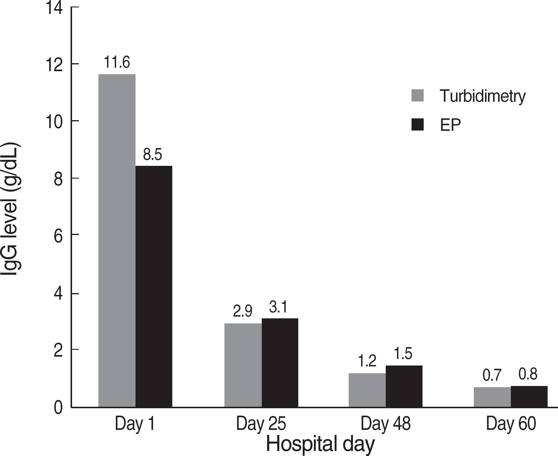
We report a case of multiple myeloma showing marked differences in serum Immunoglobulin G (IgG) levels between serum protein electrophoresis and turbidimetry. A 47-yr old man was admitted to our hospital due to severe back pain and diagnosed as having IgG-kappa type multiple myeloma. Serum protein level was 14.4 g/dL at the time of diagnosis. Serum IgG level was 8.5 g/dL by serum protein electrophoresis, but 11.6 g/dL by turbidimetry. The patient's clinical conditions had improved after receiving VAD (vincristine, adriamycin, dexamethasone) and VTD (vincristine, thalidomide, dexamethasone) chemotherapy and there were no differences in IgG levels between electrophoresis and turbidimetry when serum IgG levels were less than 3.0 g/dL. According to this, we considered that both protein electrophoresis and turbidimetry should be needed to quantify serum immunoglobulins for diagnosis and follow-up of the patients with monoclonal gammopathy.
Go to : 
REFERENCES
1.Durie BG., Salmon SE. A clinical staging system for multiple myeloma. Correlation of measured myeloma cell mass with presenting clinical features, response to treatment, and survival. Cancer. 1975. 36:842–54.

2.Greipp PR., San Miguel J., Durie BG., Crowley JJ., Barlogie B., Blade J, et al. International staging system for multiple myeloma. J Clin Oncol. 2005. 23:3412–20.

3.Katzel JA., Hari P., Vesole DH. Multiple myeloma: charging toward a bright future. CA Cancer J Clin. 2007. 57:301–18.

4.McClatchey KD, editor. Clinical Laboratory Medicine. 2nd ed.Philadelphia: Lippincott Williams & Wilkins;2002. p. 1448.
5.Kang SY., Suh JT., Lee HJ., Yoon HJ., Lee WI. Establishment of serum reference range for free light chains and its clinical usefulness in multiple myeloma. Korean J Lab Med. 2004. 24:273–8. (강소영, 서진태, 이희주, 윤휘중, 이우인. 혈청유리형경쇄 참고치 설정과 다발성골수종환자들에서의임상적의의. 대한진단검사의학회지 2004;24: 273-8.).
6.Katzmann JA., Clark RJ., Abraham RS., Bryant S., Lymp JF., Bradwell AR, et al. Serum reference intervals and diagnostic ranges for free kappa and free lambda immunoglobulin light chains: relative sensitivity for detection of monoclonal light chains. Clin Chem. 2002. 48:1437–44.
7.Bradwell AR., Carr-Smith HD., Mead GP., Tang LX., Showell PJ., Drayson MT, et al. Highly sensitive, automated immunoassay for immunoglobulin free light chains in serum and urine. Clin Chem. 2001. 47:673–80.

8.Chang CY., Fritsche HA., Glassman AB., McClure KC., Liu FJ. Underestimation of monoclonal proteins by agarose serum protein electrophoresis. Ann Clin Lab Sci. 1997. 27:123–9.
9.Katzmann JA., Massey MA., Greipp PR., Clark RJ., Thompson CK., Lust JA, et al. Artifactually low IgG monoclonal protein (M-spike) quantitation on agarose gel electrophoresis: comparison of agarose gel electrophoresis, capillary zone electrophoresis and nephelometry [Abstract 655]. AACC 52nd Annual Meeting. Clin Chem. 2000. 46:169.
Go to : 
 | Fig. 1.Results of serum protein electophoresis at the time of diagnosis (A) and on Day 48 (B). Serum levels of total protein and gamma-globulin were decreased after VAD and VTD treatment. |
 | Fig. 2.Comparison of IgG levels between turbidimetry (Iatron, Tokyo, Japan) by Hitachi 7600-110 autoanalyzer and electrophoresis (EP) according to time elapse. The discrepancy of IgG levels between two assays was remarkable on Day 1, but there were almost no differences on Day 25, 48 and 60. Total protein was 14.0 g/dL on Day 1 and decreased to 8.4, 6.4 and 6.0 g/dL on Day 25, 48 and 60, respectively. |


 PDF
PDF ePub
ePub Citation
 Print
Print


 XML Download