

 PDF
PDF ePub
ePub Citation
 Print
Print


Abstract
Background
Although cytoplasmic CD79a (cytCD79a) is a highly lineage-specific marker of B lymphoid cells and plays an important role in the diagnosis of acute leukemia, its clinical significance is not fully understood. We aimed to investigate the relationship between cytCD79a positivity and survival probability, and to evaluate the prognostic value of cytCD79a expression in AML with t(8;21) (q22;q22).
Methods
A total of 68 cases of AML with t(8;21)(q22;q22) were diagnosed based on conventional morphology, cytochemistry, flow cytometrty, and cytogenetic and molecular genetic analysis. Immuno-histochemistry of cytCD79a was performed retrospectively. Laboratory and clinical findings were reviewed.
Results
Five patients among 68 AML with t(8;21)(q22;q22) revealed cytCD79a positive reaction; scores for myeloid lineage/B-lymphoid lineage were 5/3-3.5. Among the five cytCD79a positive patients, only one patient was a child. Three patients were with refractory AML or relapsed, and two patients died within 10 months. Median survival time of cytCD79a positive group was shorter (8.0 months) than that (61.3 months) of cytCD79a negative group. The survival probability of the cytCD79a expression group was significantly lower than classical AML with t(8;21)(q22;q22) (P=0.0001).
Go to : 
REFERENCES
1.Bene MC., Castoldi G., Knapp W., Ludwig WD., Matutes E., Orfao A, et al. Proposals for the immunological classification of acute leukemias. European Group for the Immunological Characterization of Leukemias (EGIL). Leukemia. 1995. 9:1783–6.
2.Brunning RD., Head D. Acute leukaemias of ambiguous lineage. Jaffe ES, editor. World Health Organization classification of tumors. Pathology and Genetics of Tumours of Haematopoietic and Lymphoid Tissues. Washington: IARC press;2001. p. 106–7.
3.Lai R., Juco J., Lee SF., Nahirniak S., Etches WS. Flow cytometric detection of CD79a expression in T-cell acute lymphoblastic leukemias. Am J Clin Pathol. 2000. 113:823–30.

4.Engel P., Tedder TF. New CD from the B cell section of the Fifth International Workshop on Human Leukocyte Differentiation Antigens. Leuk Lymphoma. 1994. 13(S):S61–4.
5.Paredes-Aguilera R., Romero-Guzman L., Lopez-Santiago N., Burbano-Ceron L., Camacho-Del Monte O., Nieto-Martinez S. Flow cytometric analysis of cell-surface and intracellular antigens in the diagnosis of acute leukemia. Am J Hematol. 2001. 68:69–74.
6.Kozlov I., Beason K., Yu C., Hughson M. CD79a expression in acute myeloid leukemia t(8;21) and the importance of cytogenetics in the diagnosis of leukemias with immunophenotypic ambiguity. Cancer Genet Cytogenet. 2005. 163:62–7.
7.Rooney DE, Czepulkowski BH, editors. Human cytogenetics: malignancy and acquired abnormalities. A practical approach. 3rd ed.New York: Oxford university press;2001. p. 2–17.
8.Shaffer LG, Tommerup N, editors. An international system for human cytogenetic nomenclature. Basel: S. Karger;2005.
9.Sarriera JE., Albitar M., Estrov Z., Gidel C., Aboul-Nasr R., Manshouri T, et al. Comparison of outcome in acute myelogenous leukemia patients with translocation (8;21) found by standard cytogenetic analysis and patients with AML1/ETO fusion transcript found only by PCR testing. Leukemia. 2001. 15:57–61.
10.Carbonell F., Swansbury J., Min T., Matutes E., Farahat N., Buccheri V, et al. Cytogenetic findings in acute biphenotypic leukaemia. Leukemia. 1996. 10:1283–7.
11.Kim SH. Consensus for immunophenotyping of hematologic malignancy. Korean J Lab Med. 2006. 26(S2):S306–9. (김선희. Consensus for immunophenotyping of hematologic malignancy. 대한진단검사의학회지 2006;26(부록 2): S306-9.).
12.Gupta R., Bain BJ., Knight CL. Cytogenetic and molecular genetic abnormalities in systemic mastocytosis. Acta Haematol. 2002. 107:123–8.
13.Swolin B., Rodjer S., Roupe G. Cytogenetic studies in patients with mastocytosis. Cancer Genet Cytogenet. 2000. 120:131–5.
14.Killick S., Powles RL., Hamblin M., Treleaven JG., Zomas A., Matutes E, et al. Outcome of biphenotypic acute leukemia. Haematologica. 1999. 84:699–706.
15.Scolnik MP., Aranguren PN., Cuello MT., Palacios MF., Sanjurjo J., Bracco MM, et al. Biphenotypic acute leukemia with t(15;17). Leuk Lymphoma. 2005. 46:607–10.
16.He G., Wu D., Sun A., Xue Y., Jin Z., Qiu H, et al. CytCD79a expression in acute leukemia with t(8;21): biphenotypic or myeloid leukemia? Cancer Genet Cytogenet. 2007. 174:76–7.
17.Tiacci E., Pileri S., Orleth A., Pacini R., Tabarrini A., Frenguelli F, et al. PAX5 expression in acute leukemias: higher B-lineage specificity than CD79a and selective association with t(8;21)-acute myelogenous leukemia. Cancer Res. 2004. 64:7399–404.
18.Konoplev S., Bueso-Ramos CE. Advances in the pathologic diagnosis and biology of acute myeloid leukemia. Ann Diagn Pathol. 2006. 10:39–65.
19.Bene MC. Immunophenotyping of acute leukaemias. Immunol Lett. 2005. 98:9–21.
20.Yoo SJ., Chi HS., Jang S., Seo EJ., Seo JJ., Lee JH, et al. Quantification of AML1-ETO fusion transcript as a prognostic indicator in acute myeloid leukemia. Haematologica. 2005. 90:1493–501.
21.Miyamoto T., Nagafuji K., Harada M., Niho Y. Significance of quantitative analysis of AML1/ETO transcripts in peripheral blood stem cells from t(8;21) acute myelogenous leukemia. Leuk Lymphoma. 1997. 25:69–75.
22.Nimer SD., Moore MA. Effects of the leukemia-associated AML1-ETO protein on hematopoietic stem and progenitor cells. Oncogene. 2004. 23:4249–54.
23.The value of c-kit in the diagnosis of biphenotypic acute leukemia. EGIL (European Group for the Immunological Classification of Leukaemias). Leukemia. 1998. 12:2038.
24.Lee ST., Kim HJ., Kim SH. Defining an optimal number of immunophenotypic markers for lineage assignment of acute leukemias based on the EGIL scoring system. Korean J Lab Med. 2006. 26:393–9. (이승태, 김희진, 김선희. 급성백혈병의 lineage assignment에있어서면역표현형표지자의적정개수. 대한진단검사의학회지 2006;26: 393-9.).
Go to :
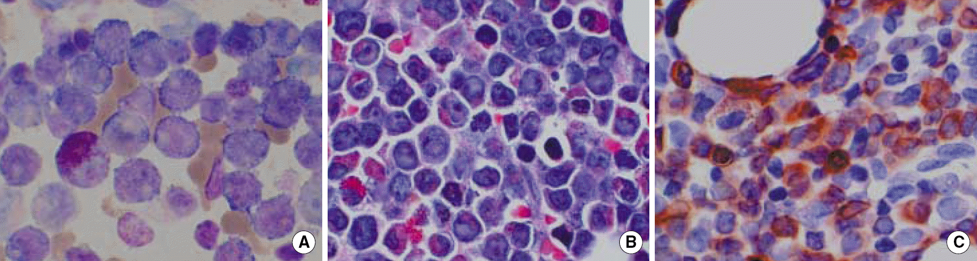 | Fig. 1.Bone marrow findings of a patient in the cytCD79a expression group, ×1,000. (A: aspiration, Wright stain; B: biopsy, hematoxylin and eosin stain; C: biopsy, cytCD79a immunohistochemistry). Abbreviation: cytCD79a, cytoplasmic CD79a. |
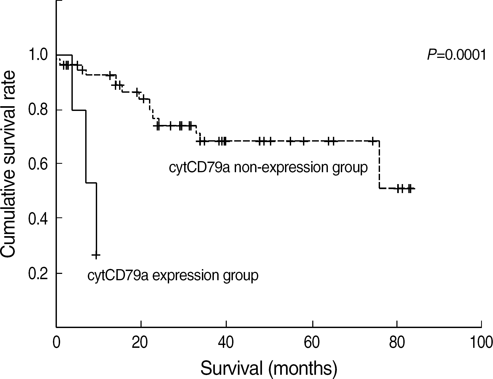 | Fig. 2.The survival probability of the cytCD79a expression group was significantly lower than the AML with t(8;21). Abbreviation: See Fig.1. |
Table 1.
Scoring system for the definition of BAL proposed by European Group for the Immunologic Classification of Leukemia (EGIL)
| Score | B lineage | T lineage | Myeloid lineage |
|---|---|---|---|
| 2 | cytCD79a | CD3 (m/cyt) | MPO∗ |
| cyt IgM | Anti-TCR | ||
| cytCD22 | |||
| 1 | CD19 | CD2 | CD117 |
| CD20 | CD5 | CD13 | |
| CD10 | CD8 | CD33 | |
| CD10 | CD65 | ||
| 0.5 | TdT | TdT | CD14 |
| CD24 | CD7 | CD15 | |
| CD1a | CD64 |
Table 2.
Characteristics of the five patients showing cytCD79a positivity in immunohistochemistry
| UPN | Age | Sex | FAB subtype | Blast (%) | Cellularity (%) | Myeloid | B lymphoid | Karyotype | AML1/ETO rearrangement∗ | ||||||||
|---|---|---|---|---|---|---|---|---|---|---|---|---|---|---|---|---|---|
| MPO | CD13 | CD33 | CD117 | Score | cytC D79a | CD19 | CD10 | TdT | Score | ||||||||
| 1 | 5 | F | M4 | 44.8 | 70 | + | + | + | + | 5 | + | + | - | + | 3.5 | 45,X,-X,t(8;21)(q22;q22)[20] | + |
| 2 | 38 | F | M1 | 25.4 | 95 | + | + | + | + | 5 | + | + | - | - | 3 | 46,XX,t(8;21)(q22;q22)[20] | + |
| 3 | 54 | M | M2 | 68.2 | 80 | + | + | + | + | 5 | + | + | - | - | 3 | 45,X,-Y,t(8;21)(q22;q22)[20] | + |
| 4 | 49 | M | M2 | 46.4 | 95 | + | + | + | + | 5 | + | + | - | - | 3 | 45,X,-Y,t(8;21)(q22;q22)[20] | + |
| 5 | 45 | F | M2 | 57.2 | 100 | + | + | + | + | 5 | + | + | - | - | 3 | 46,XX,t(8;21)(q22;q22), add(11)(p11.2)[20] | + |
Table 3.
Clinical and laboratory findings according to cytCD79a expression on leukemic cells
| Expression of cytCD79a (n=68) | P value | ||
|---|---|---|---|
| Positive (n=5) | Negative (n=63) | ||
| Median age (yr) | 49 (5-58) | 36 (3-73) | 0.466 |
| Sex (M:F) | 2:3 | 28:35 | 0.528 |
| FAB subtype | M1, 2, 4 | M1, 2, 4 | |
| Leukemic cells in BM (%)∗ | 59.2±16.9 | 60.1±22.6 | 0.174 |
| Cellularity (%)∗ | 88.0±12.6 | 81.0±21.7 | 0.180 |
| WBC count (μ/L) | 46,190±6,860 | 29,920±5,610 | 0.070 |
| BM fibrosis (%) | 20 | 14.3 | 0.857 |
| Immunophenotyping (%) | |||
| TdT | 20.0 (1) | 25.4 (16) | 0.399 |
| CD34 | 80.0 (4) | 87.3 (55) | 0.926 |
| CD19 | 100.0 (5) | 52.4 (33) | 0.048 |
| Prognosis | |||
| Relapse (%) | 3 (60.0) | 16 (25.4) | |
| Mortality (%) | 2 (40.0) | 14 (22.2) | |
| Median survival (months) | 8.0 | 61.3 | <0.001 |
 XML Download