Abstract
Background
HLA-DR negativity is known to be useful for distinguishing acute promyelocytic leukemia (APL) from other subtypes of AML, but non-APL cases without HLA-DR antigen expression have been reported. The purpose of this study was to evaluate and compare the characteristics of APL, HLA-DR negative non-APL, and HLA-DR positive non-APL cases.
Methods
A total of 114 cases of AML admitted at Ewha Womans University, Mokdong Hospital between March 1997 and June 2006 were included in this study. A diagnosis of AML was made based on the results of morphology, cytochemistry, immunophenotype, cytogenetics, and/or fluorescence in situ hybridization.
Results
Among the 114 AML patients, HLA-DR antigen was not expressed in 39 (34%), including 24 non-APL (62%) and 15 APL patients (38%). The HLA-DR negative non-APL group showed higher leukocyte counts and positive rate of CD19 expression than did APL group (P<0.05). The remaining laboratory findings were not statistically different between the HLA-DR negative non-APL and APL groups. CD34 expression was more frequent in the HLA-DR positive non-APL group than in the HLA-DR negative non-APL group and APL group. Of the 24 patients with HLA-DR negative non-APL, 7 patients had disseminated intravascular coagulation and 2 patients showed morphologic features similar to those of APL.
REFERENCES
1.Bene MC. Immunophenotyping of acute leukaemias. Immunol Lett. 2005. 98:9–21.
2.Casasnovas RO., Slimane FK., Garand R., Faure GC., Campos L., Deneys V, et al. Immunological classification of acute myeloblastic leukemias: relevance to patient outcome. Leukemia. 2003. 17:515–27.

3.Bene MC., Bernier M., Castoldi G., Faure GC., Knapp W., Ludwig WD, et al. Impact of immunophenotyping on management of acute leukemias. Haematologica. 1999. 84:1024–34.
4.Kaleem Z., Crawford E., Pathan MH., Jasper L., Covinsky MA., Johnson LR, et al. Flow cytometric analysis of acute leukemias. Diagnostic utility and critical analysis of data. Arch Pathol Lab Med. 2003. 127:42–8.
5.Jennings CD., Foon KA. Recent advances in flow cytometry: application to the diagnosis of hematologic malignancy. Blood. 1997. 90:2863–92.

6.Paietta E., Andersen J., Gallagher R., Bennett J., Yunis J., Cassileth P, et al. The immunophenotype of acute promyelocytic leukemia (APL): an ECOG study. Leukemia. 1994. 8:1108–12.
7.Oelschlagel U., Nowak R., Mohr B., Thiede C., Ehninger G., Schaub A, et al. Specificity of immunophenotyping in acute promyelocytic leukemia. Cytometry. 2000. 42:396–7.
8.Exner M., Thalhammer R., Kapiotis S., Mitterbauer G., Knobl P., Haas OA, et al. The “typical” immunophenotype of acute promyelocytic leukemia (APL-M3): does it prove true for the M3-variant? Cytometry. 2000. 42:106–9.

9.Baer MR., Stewart CC., Dodge RK., Leget G., Sule N., Mrozek K, et al. High frequency of immunophenotype changes in acute myeloid leukemia at relapse: implications for residual disease detection (Cancer and Leukemia Group B Study 8361). Blood. 2001. 97:3574–80.

10.Wetzler M., McElwain BK., Stewart CC., Blumenson L., Mortazavi A., Ford LA, et al. HLA-DR antigen-negative acute myeloid leukemia. Leukemia. 2003. 17:707–15.

11.Orfao A., Chillon MC., Bortoluci AM., Lopez-Berges MC., Garcia-Sanz R., Gonzalez M, et al. The flow cytometric pattern of CD34, CD15 and CD13 expression in acute myeloblastic leukemia is highly characteristic of the presence of PML-RARalpha gene rearrangements. Haematologica. 1999. 84:405–12.
12.Fenu S., Carmini D., Mancini F., Guglielmi C., Alimena G., Riccioni R, et al. Acute myeloid leukemias M2 potentially misdiagnosed as M3 variant French-American-Britain (FAB) subtype: a transitional form? Leuk Lymphoma. 1995. 18(S1):49–55.

13.Lazarchick J., Hopkins M. HLA-Dr negative acute non-lymphocytic leukemia. Ann Clin Lab Sci. 1998. 28:150–2.
14.Jaffe ES HN., Stein H, et al. WHO classification of tumours: Tumours of Hematopoietic and Lymphoid tissue. Lyon: IARC Press;2001.
15.Bene MC., Castoldi G., Knapp W., Ludwig WD., Matutes E., Orfao A, et al. Proposals for the immunological classification of acute leukemias. European Group for the Immunological Characterization of Leukemias (EGIL). Leukemia. 1995. 9:1783–6.
16.Guglielmi C., Martelli MP., Diverio D., Fenu S., Vegna ML., Cantu-Rajnoldi A, et al. Immunophenotype of adult and childhood acute promyelocytic leukaemia: correlation with morphology, type of PML gene breakpoint and clinical outcome. A cooperative Italian study on 196 cases. Br J Haematol. 1998. 102:1035–41.

17.Khoury H., Dalal BI., Nantel SH., Horsman DE., Lavoie JC., Shepherd JD, et al. Correlation between karyotype and quantitative immunophenotype in acute myelogenous leukemia with t(8;21). Mod Pathol. 2004. 17:1211–6.

18.Syampurnawati M., Tatsumi E., Furuta K., Takenokuchi M., Nakamachi Y., Kawano S, et al. HLA-DR-negative AML (M1 and M2): FLT3 mutations (ITD and D835) and cell-surface antigen expression. Leuk Res. 2007. 31:921–9.
19.Kussick SJ., Stirewalt DL., Yi HS., Sheets KM., Pogosova-Agadjanyan E., Braswell S, et al. A distinctive nuclear morphology in acute myeloid leukemia is strongly associated with loss of HLA-DR expression and FLT3 internal tandem duplication. Leukemia. 2004. 18:1591–8.
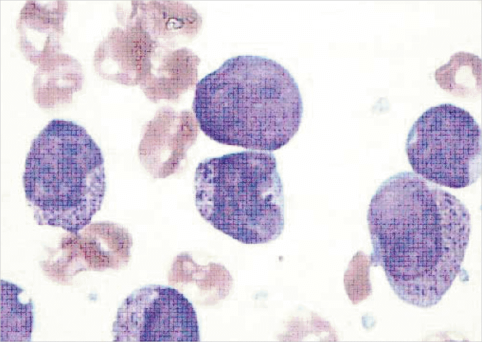
Fig. 1.
A case of 21 yr-old female patient diagnosed as HLA-DR negative AML. Immature cells showed indented nuclear margins and coarse cytoplasmic granules (BM aspiration, Wright stain, ×1,000).

Table 1.
Comparison of the characteristics of non-APL and APL according to HLA-DR expression
| APL (N=16) | HLA-DR negative non APL (N=24) | HLA-DR positive non APL (N=74) | |
|---|---|---|---|
| Age (yr), median (range) | 36 (16-80) | 46 (4-68) | 49 (5-81) |
| Sex (M:F) | 9:7 | 11:13 | 38:36 |
| WBC (×109/L)∗, median (range) | 2.2 (0.4-76.2) | 47.4 (1-497.6) | 13.1 (0.3-554.4) |
| BM leukemic cell (%)†, median (range) | 74 (47.6-97.8) | 89.8 (21.4-99.2) | 67.1 (22.0-98.8) |
| Hb (g/dL), median (range) | 7.7 (4.7-11.2) | 8.5 (5.7-12.0) | 8.1 (4.6-14.9) |
| PLT (×109/L)†, median(range) | 32 (10-104) | 37 (1-177) | 50 (4-538) |
| DIC (%)† | 7/15 (46.7) | 7/18 (38.9) | 5/44 (11.3) |
| FAB classification (%) | 2 (8) | 5 (7) | |
| M0 | |||
| M1 | 16 (67) | 20 (27) | |
| M2 | 4 (17) | 36 (49) | |
| M3 | 16 (100) | ||
| M4 | 1 (4) | 11 (15) | |
| M5 | 0 (0) | 0 (0) | |
| M6 | 1 (1) | ||
| Others‡ | 1 (4) | 1 (1) | |
| Positive of antigen expression (%) | |||
| CD 13 | 15 (94) | 18 (75) | 66 (89) |
| CD 33 | 16 (100) | 22 (92) | 71 (96) |
| HLA-DR† | 1 (6) | 0 (0) | 74 (100) |
| CD 34† | 1 (6) | 5 (21) | 52 (70) |
| CD 7 | 1 (6) | 1 (4) | 13 (18) |
| CD 5 | 0 (0) | 0 (0) | 1 (1) |
| CD 3 | 0 (0) | 0 (0) | 2 (3) |
| CD 19∗ | 1 (6) | 11 (46) | 23 (31) |
| CD 10 | 0 (0) | 0 (0) | 1 (1) |
| CD 22 | 0 (0) | 1 (4) | 6 (8) |
| CD 20 | 0 (0) | 0 (0) | 2 (3) |
| CD 14 | 0 (0) | 1 (4) | 10 (14) |
| CD 16/56 | 0 (0) | 3 (13) | 7 (9) |
| Karyotype (%) | |||
| t(8;21)(q22;q22) | 0 (0) | 8 (12) | |
| t(15;17)(q22;q21) | 16 (100) | 0 (0) | 0 (0) |
| inv(16) | 0 (0) | 2 (3) | |
| t(9;22)(q34;q11) | 0 (0) | 3 (5) | |
| Other abnormalities | 2 (12) | 14 (22) | |
| Normal karyotype | 14 (88) | 38 (58) | |
| Not available | 8 | 9 |


 PDF
PDF ePub
ePub Citation
 Print
Print


 XML Download