

 PDF
PDF ePub
ePub Citation
 Print
Print


Abstract
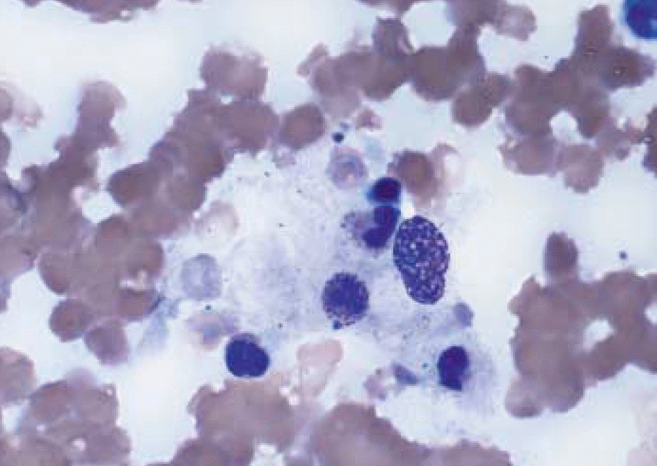
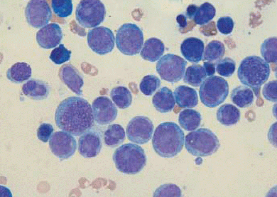
We report a case of therapy-related acute myeloid leukemia after low-dosed topoisomerase II inhibitor (etoposide) treatment for hemophagocytic lymphohistiocytosis (HLH). A 62-yr-old female patient had previously been treated with a HLH-94 protocol containing a low-dose of etoposide (total dose of 300 mg/m2). Thirty-one months later, the patient was admitted to the hematology department with general weakness and upper respiratory infection symptoms. Peripheral blood smear and bone marrow study revealed acute monocytic leukemia. There was no evidence of myelodysplastic syndrome, and a cytogenetic study showed no chromosomal abnormalities.
References
1. Verbsky JW, Grossman WJ. Hemophagocytic lymphohistiocytosis: diagnosis, pathophysiology, treatment, and future perspectives. Ann Med. 2006; 38:20–31.

2. Chang SS. Hemophagocytic lymphohistiocytosis. Korean J Lab Med. 2006; 26(S1):S136–8.
3. Kim KH, Kim SH, Lee JK, Cho YJ, Kim YK, Shin DH, et al. A case of Epstein-barr virus-associated hemophagocytic lymphohistiocytosis with clonal karyotype abnormality. Korean J Lab Med. 2005; 25:85–9.
4. Lee JS, Kang JH, Lee GK, Park HJ. Successful treatment of Epstein-Barr virus-associated hemophagocytic lymphohistiocytosis with HLH-94 protocol. J Korean Med Sci. 2005; 20:209–14.
5. Cho HS, Park YN, Lyu CJ, Park SM, Oh SH, Yang CH, et al. EBV-elicited familial hemophagocytic lymphohistiocytosis. Yonsei Med J. 1997; 38:245–8.
6. Janka GE, Schneider EM. Modern management of children with haemophagocytic lymphohistiocytosis. Br J Haematol. 2004; 124:4–14.
7. Imashuku S, Teramura T, Kuriyama K, Kitazawa J, Ito E, Morimoto A, et al. Risk of etoposide-related acute myeloid leukemia in the treatment of Epstein-Barr virus-associated hemophagocytic lymphohistiocytosis. Int J Hematol. 2002; 75:174–7.
8. Kitazawa J, Ito E, Arai K, Yokoyama M, Fukayama M, Imashuku S. Secondary acute myelocytic leukemia after successful chemotherapy with etoposide for Epstein-Barr virus-associated hemophagocytic lymphohistiocytosis. Med Pediatr Oncol. 2001; 37:153–4.
9. Jaffe ES, Harris NL, editors. Pathology and genetics of tumours of haematopoietic and lymphoid tissues-World health organization classification of tumours. Lyon: International Agency for Research on Cancer;2001. p. 89–91.
10. Goto H, Shimazaki C, Tatsumi T, Yamagata N, Inaba T, Fujita N, et al. Acute myelomonocytic leukemia after treatment with chronic oral etoposide: are MLL and LTG9 genes targets for etoposide? Int J Hematol. 1994; 60:145–9.
11. Nakamura H, Ishizaki T, Itoyama T, Soda H, Yoshida Y, Yamada Y, et al. Acute myeloid leukaemia with t(9;11)(p22;q23) in a patient treated for adult T cell leukaemia. Br J Haematol. 1994; 86:222–4.
12. Stine KC, Saylors RL, Sawyer JR, Becton DL. Secondary acute myelogenous leukemia following safe exposure to etoposide. J Clin Oncol. 1997; 15:1583–6.
13. Silva ML, Land MG, Maradei S, Otero L, Veith M, Brito G, et al. Translocation (11;11)(p13-p15;q23) in a child with therapy-related acute myeloid leukemia following chemotherapy with DNA-topoisomerase II inhibitors for Langerhans cell histocytosis. Cancer Genet Cytogenet. 2002; 135:101–2.
14. Takahashi T, Yagasaki F, Endo K, Takahashi M, Itoh Y, Kawai N, et al. Therapy-related AML after successful chemotherapy with low dose etoposide for virus-associated hemophagocytic syndrome. Int J Hematol. 1998; 68:333–6.
15. Suzuki T, Mugishima H, Yamada A, Nagata T, Shichino H, Chin M, et al. Development of acute lymphoblastic leukemia following hemophagocytic lymphohistiocytosis: is it secondary leukemia? Int J Hematol. 1999; 70:58–9.
 XML Download