

 PDF
PDF ePub
ePub Citation
 Print
Print


Abstract
The inv(16)(p13q22) is found in de novo AML and is closely associated with the FAB subtype M4eo. The inv(16) is rarely reported in therapy-related AML (t-AML) patients. Herein, we report a case of t-AML with inv(16) after combination chemotherapy using antimitotic agent and alkylating agent (cis-platin-paclitaxel) for ovarian serous cystadenocarcinoma.
References
1. Jaffe ES, Harris NL, editors. World Health Organization classification of tumors. Pathology and genetics of tumors of haematopoietic and lymphoid tissue. France: IARC Press, Lyon;2001. p. 82–4. 89–91.
2. Andersen MK, Larson RA, Mauritzson N, Schnittger S, Jhanwar SC, Pederson-Bjergaard J. Balanced chromosome abnormalities inv(16) and t(15;17) in therapy-related myelodysplastic syndromes and acute leukemia: report from an international workshop. Genes Chromosomes Cancer. 2002; 33:395–400.

3. Yoon T, Kim DY, Lee KW, Kim DH, Yoon SS, Park SY, et al. A case of therapy-related acute lymphoblastic leukemia after 131 I-treatment for thyroid papillary carcinoma. Korean J Med. 2004; 66:437–41.
4. Song SH, Bin JS, Kim JH, Park YS, Park KC, Shun DJ, et al. Therapy-related acute myelogenous leukemia with complex chromosomal defect. Report of a case. Korean J Hematol. 1992; 27:117–22.
5. Kim M, Lim J, Kim Y, Han K, Kang CS, Kim HJ, et al. A case of therapy-related acute myeloid leukemia associated with inv(16), with subsequent development of t(9;22). Leukemia. 2006; 20:746–8.
6. Armitage JO, Carbone PP, Connors JM, Levine A, Bennett JM, Kroll S. Treatment-related myelodysplasia and acute leukemia in non-Hodgkin's lymphoma patients. J Clin Oncol. 2003; 21:897–906.
7. Dissing M, Le Beau MM, Pedersen-Bjergaard J. Inversion of chromosome 16 and uncommon rearrangements of the CBFB and MYH11 genes in therapy-related acute myeloid leukemia: rare events related to DNA-topoisomerase II inhibitors? J Clin Oncol. 1998; 16:1890–6.
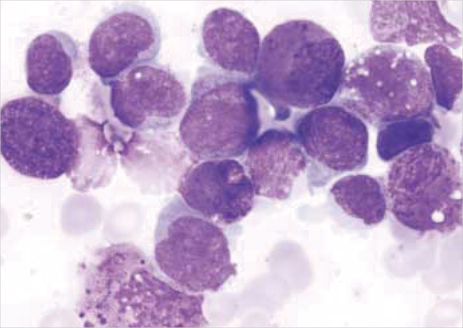
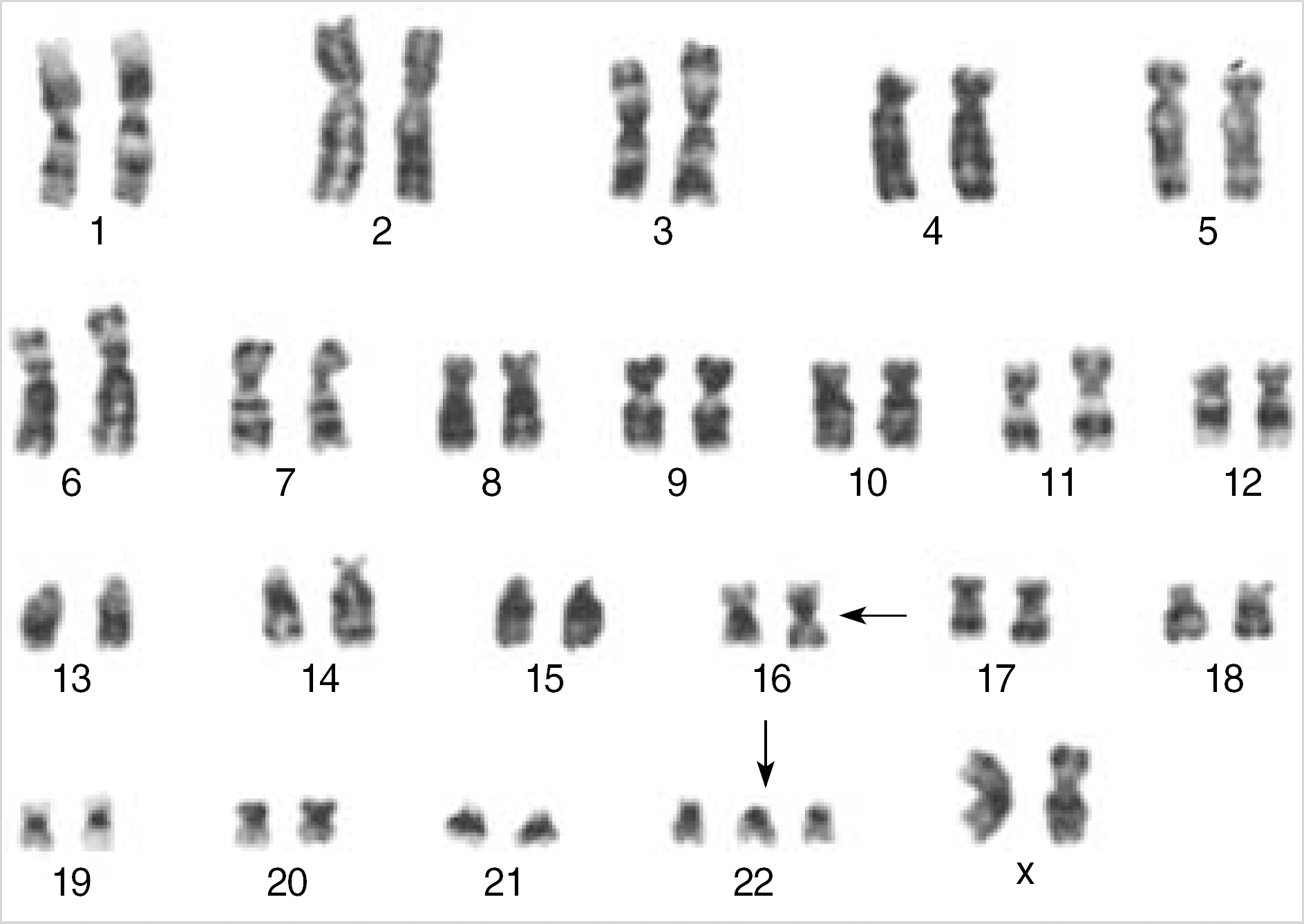
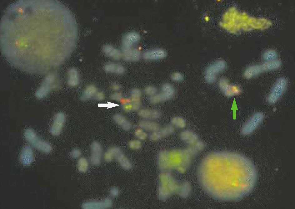
Fig. 3.
FISH analysis with LSI 5′ CBFβ (Red)/ LSI 3′ CBFβ (Green) dual color, break apart rearrangement probe (Vysis, Inc). Normal chromosome 16 showed overlapping red/green signals (green arrow) and abnormal chromosome 16 split the CBFβ locus resulting in separate red and green signals (white arrow).
 XML Download