

 PDF
PDF ePub
ePub Citation
 Print
Print


Abstract
Background
Fibrin-ring granuloma (FRG), which can be found in bone marrow or the liver, is a subtype of epithelioid granuloma characterized by a central fat vacuole and annular peripheral fibrinoid materials. FRG has been proven to be associated with many etiologies such as several infectious organisms (Coxiella burnett; Epstein-Barr Virus, EBV; cytomegalovirus, CMV; and hepatitis A virus), allopurinol induced hepatitis, Hodgkin's lymphoma, and peripheral T-cell lymphoma.
Methods
We retrospectively reviewed 24 patients diagnosed with FRG by bone marrow biopsy at a single institute between 1995 and 2004. We reviewed clinical symptoms and laboratory findings of the patients, classified them by etiology, and compared prognosis of each group.
Results
The most common cause of FRG was acute or chronic EBV infection. Chronic or acute EBV infection was associated with 41.4% of patients (10/24). Of the remaining patients, 33.3% (8/24) were leukemia or lymphoma patients after chemotherapy, 4.2% (1/24) was a patient with hepatic failure, and 20.8% (5/24) were diagnosed as fever of unknown origin. The most common symptom and clinical finding were fever and cytopenia. EBV-associated group comprised chronic active EBV infection, EBV-associated hemophagocytic histiocytosis, acute EBV infection, EBV-associated lymphoproliferative disease, and Langerhans' cell histiocytosis. The EBV-associated group showed a lower survival probability compared with the non-EBV group (P<0.05).
Go to : 
References
1. Blanco P, Viallard JF, Parrens M, Mercie P, Pellegrin JL. Bone-marrow fibrin-ring granuloma. Lancet. 2003; 362:1224.

2. Kvasnicka HM, Thiele J. Differentiation of granulomatous lesions in the bone marrow. Pathologe. 2002; 23:465–71.
3. Okun DB, Sun NC, Tanaka KR. Bone marrow granulomas in Q fever. Am J Clin Pathol. 1979; 71:117–21.
4. Voigt JJ, Delsol G, Fabre J. Liver and bone marrow granulomas in Q fever. Gastroenterology. 1983; 84:887–8.
5. Young JF, Goulian M. Bone marrow fibrin ring granulomas and cytomegalovirus infection. Am J Clin Pathol. 1993; 99:65–8.
6. Vanderstigel M, Zafrani ES, Lejonc JL, Schaeffer A, Portos JL. Allopurinol hypersensitivity syndrome as a cause of hepatic fibrin-ring granulomas. Gastroenterology. 1986; 90:188–90.
7. Raya Sanchez JM, Arguelles HA, Brito Barroso ML, Nieto LH. Bone marrow fibrin-ring (doughnut) granulomas and peripheral T-cell lymphoma: an exceptional association. Haematologica. 2001; 86:112.
8. Niesters HG, van Esser J, Fries E, Wolthers KC, Cornelissen J, Osterhaus AD. Development of a real-time quantitative assay for detection of Epstein-Barr virus. J Clin Microbiol. 2000; 38:712–5.
9. Ende N, Gelpi AP. Pathologic changes noted in bone marrow in a case of Q fever. AMA Arch Internal Med. 1957; 100:793–6.
10. Ruel M, Sevestre H, Henry-Biabaud E, Courouce AM, Capron JP, Erlinger S. Fibrin ring granulomas in hepatitis A. Dig Dis Sci. 1992; 37:1915–7.
11. Nenert M, Mavier P, Dubuc N, Deforges L, Zafrani ES. Epstein-Barr virus infection and hepatic fibrin-ring granulomas. Hum Pathol. 1988; 19:608–10.
12. Cohen JI. Epstein-Barr virus infection. N Engl J Med. 2000; 343:481–92.
13. Oh SH, Lee YA, Moon WY, Ko TS, Park YS, Moon HN, et al. Prevalence of Epstein-Barr Vvirus (EBV) antibody in Korean children. J Korean Pediatr Soc. 1994; 37:804–11.
14. Fiala M, Colodro I, Talbert W, Ellis R, Chatterjee S. Bone marrow granulomas in mononucleosis. Postgrad Med J. 1987; 63:277–9.
15. Straus SE. The chronic mononucleosis syndrome. J Infect Dis. 1988; 157:405–12.
16. Kang ES, Gong GY, Chi HS. A case of fibrin-ring granuloma in bone marrow and liver associated with Epstein-Barr virus infection. Korean J Clin Pathol. 1995; 15:178–82.
17. de Bayser L, Roblot P, Ramassamy A, Silvain C, Levillain P, Becq-Giraudon B. Hepatic fibrin-ring granulomas in giant cell arteritis. Gastroenterology. 1993; 105:272–3.
18. Delsol G, Pellegrin M, Voigt JJ, Fabre J. Diagnostic value of granuloma with fibrinoid ring. Am J Clin Pathol. 1980; 73:289.
19. Rexroth G, Rosch W, Altmannsberger M. Bone marrow granulomatosis in Q-fever. Med Klin (Munich). 2000; 95:404–8.
20. Srigley JR, Vellend H, Palmer N, Phillips MJ, Geddie WR, Van Nostrand AW, et al. Q-fever. The liver and bone marrow pathology. Am J Surg Pathol. 1985; 9:752–8.
Go to :
 | Fig. 1.(A) Multiple fibrin-ring granulomas in UPN 5 (BM clot section, Hematoxylin-Eosin stain, ×400). EBV PCR with BM sample revealed positive. (B) Fibrin-ring granulomas in UPN 10. Serial result of real-time EBV quantitative PCR presented 15,720 copies/mL at the diagnosis of FRG and decreased to 2,490 copies/mL after 10 days (BM biopsy specimen, Hematoxylin-Eosin stain, ×400). (C) Masson's trichrome stain of fibrin-ring granulomas in UPN 10 (BM biopsy specimen, trichrome stain, ×400). |
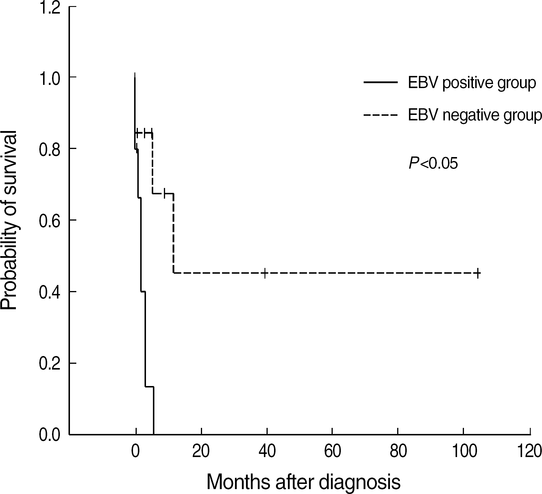 | Fig. 2.Kaplan-Meier survival curves of patients. EBV positive group shows a significantly low survival rate. The median survival of EBV positive group is 1.4 months (mean=2.0 months) and that of negative group is 11.8 months (mean=50.7 months).
Abbreviation: UPN, unique patient number.
|
Table 1.
Clinical and laboratory data of patients
Abbreviations: UPN, unique patient number; LAP, lymphadenopathry; NT, not tested; CAEBV, chronic active EBV infection; EBV-HLH, EBV associated hemophagocytic lymphohistiocytosis; LCH, Langerhans' cell histiocytosis; LPD, Lymphoproliferative disease; ISH, in situ hybridization; FUO, fever of unknown origin.
Table 2.
Four groups of patients with fibrin-granuloma according to their etiology
| Group | N of patient (n) | % of patient |
|---|---|---|
| I Acute or Chronic EBV infection | 10 | 41.7 |
| II Leukemia or lymphoma after chemotherapy | 8 | 33.3 |
| III Hepatic failure | 1 | 4.2 |
| IV FUO | 5 | 20.8 |
| Total | 24 | 100.0 |
Table 3.
Review of the literature: bone marrow fibrin-ring granuloma
 XML Download