

 PDF
PDF ePub
ePub Citation
 Print
Print


Abstract
Background
Macrocytic anemias are commonly seen in clinical practice, and precise etiologic diagnosis is essential for proper management. We evaluated the clinical utility of reticulocyte maturation parameters in macrocytic anemias to discriminate among myelodysplastic syndrome (MDS), megaloblastic anemia (MA), and non-megaloblastic macrocytic anemia associated with chronic liver disease (MA-CLD).
Methods
Using an automated reticulocyte counter, we retrospectively analyzed and compared reticulocyte maturation parameters including immature reticulocyte fraction (IRF), mean reticulocyte volume (MRV), mean sphered cell volume (MSCV) of normal control (N=34), and patients diagnosed with MDS (N=31), MA (N=52), and MA-CLD (N=196).
Results
Macrocytic anemias from MA, MDS and MA-CLD showed higher values of reticulocyte maturation parameters including IRF, MRV and MSCV than normal control (P<0.01). MDS showed higher values of reticulocyte maturation parameters including IRF, MRV and MSCV than MA-CLD (P<0.01). IRF and MRV were significantly lower in MA-CLD than in both MA and MDS (P<0.01). MSCV was significantly higher in MDS than in MA (P<0.01).
Conclusions
This study indicates that the measurement of reticulocyte maturation parameters may be a useful tool in the differential diagnosis of macrocytic anemia. The presence of high values of IRF (≥0.39), MRV (≥ 129.5 fL), and MSCV (≥ 102.3 fL) makes the diagnosis of MA-CLD unlikely and underlying MDS should be considered.
References
1. Colon-Otero G, Menke D, Hook CC. A practical approach to the differential diagnosis and evaluation of the adult patient with macrocytic anemia. Med Clin North Am. 1992; 76:581–97.

2. Breedveld FC, Bieger R, van Wermeskerken RK. The clinical significance of macrocytosis. Acta Med Scand. 1981; 209:319–22.
3. Davidson RJ, Hamilton PJ. High mean red cell volume: its incidence and significance in routine haematology. J Clin Pathol. 1978; 31:493–8.
4. Wymer A, Becker DM. Recognition and evaluation of red blood cell macrocytosis in the primary care setting. J Gen Intern Med. 1990; 5:192–7.
5. Hoffbrand V, Provan D. ABC of clinical haematology. Macrocytic anaemias. BMJ. 1997; 314:430–3.
6. Parker JE, Mufti GJ. Ineffective haematopoiesis and apoptosis in myelodysplastic syndromes. Br J Haematol. 1998; 101:220–30.
7. Park KH, Lee YK, Choi TY, Kim WB, Lee DW. Clinical significance of immature reticulocyte fraction determined by automated blood cell analyzer. Korean J Hematol. 1999; 34:281–7.
8. Hong HR, Chae SL, Park AJ. A study of reticulocyte mean channel fluorescence for differential diagnosis of anemia. Korean J Clin Pathol. 1996; 16:12–9.
9. Choi JW, Pai SH. Influence of iron depletion on immature reticulocyte fractions and reticulocyte maturity index. J Clin Pathol & Quality Control. 2000; 22:235–41.
10. Bae HG, Heo WB, Lee NY, Suh JS. Clinical utility of reticulocyte parameters in the early detection of hematopoietic engraftment after stem cell transplantation. Korean J Lab Med. 2003; 23:299–303.
11. Torres Gomez A, Casano J, Sanchez J, Madrigal E, Blanco F, Alvarez MA. Utility of reticulocyte maturation parameters in the differential diagnosis of macrocytic anemias. Clin Lab Haematol. 2003; 25:283–8.
12. Chang CC, Kass L. Clinical significance of immature reticulocyte fraction determined by automated reticulocyte counting. Am J Clin Pathol. 1997; 108:69–73.
13. Brugnara C. Use of reticulocyte cellular indices in the diagnosis and treatment of hematologic disorders. Int J Clin Lab Res. 1998; 28:1–11.
14. Watanabe K, Kawai Y, Takeuchi K, Shimizu N, Iri H, Ikeda Y, et al. Reticulocyte maturity as an indicator for estimating qualitative abnormality of erythropoiesis. J Clin Pathol. 1994; 47:736–9.
15. Daliphard S, Bizet M, Callat MP, Beufe S, Latouche JB, Soufiani H, et al. Evaluation of reticulocyte subtype distribution in myelodysplastic syndromes. Am J Hematol. 1993; 44:210–1.
16. Bowen D, Williams K, Phillips I, Cavill I. Cytometric analysis and maturation characteristics of reticulocytes from myelodysplastic patients. Clin Lab Haematol. 1996; 18:155–60.
17. Chiron M, Cynober T, Mielot F, Tchernia G, Croisille L. The GEN.S: a fortuitous finding of a routine screening test for hereditary spherocytosis. Hematol Cell Ther. 1999; 41:113–6.
18. Banfi G, Mauri C, Morelli B, Di Gaetano N, Malgeri U, Melegati G. Reticulocyte count, mean reticulocyte volume, immature reticulocyte fraction, and mean sphered cell volume in elite athletes: reference values and comparison with the general population. Clin Chem Lab Med. 2006; 44:616–22.
Fig. 1.
Reticulocyte maturation parameters. When MDS and MA-CLD are compared, there are statistical differences (P<0.01) in all reticulocyte maturation parameters: IRF, MRV and MSCV.
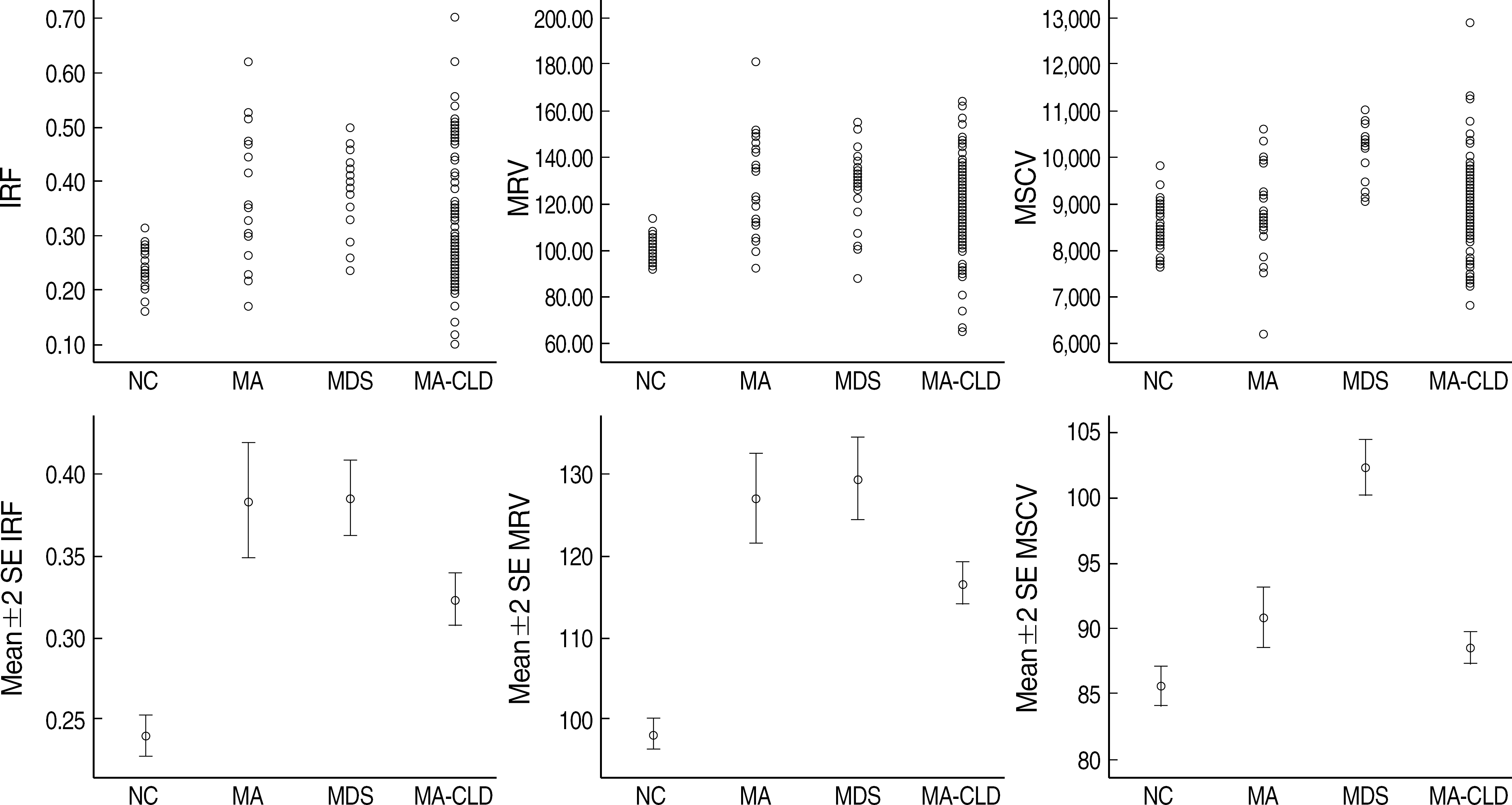
Table 1.
Routine hematological parameters
| Parameters |
Mean (SD) |
|||
|---|---|---|---|---|
| NC (N=34) | MA (N=52) | MDS (N=31) | MA-CLD (N=196) | |
| Hemoglobin (g/dL) | 14.38 (1.04) | 10.06 (1.34) | 9.30 (1.85) | 10.09 (20.3) |
| Hematocrit (%) | 41.14 (2.78) | 28.81 (3.89) | 26.85 (5.35) | 29.23 (4.74) |
| MCV (fL) | 89.56 (3.60) | 104.06 (7.06) | 101.11 (6.62) | 102.76 (4.24) |
| MCH (pg) | 31.30 (1.32) | 36.33 (2.59) | 35.02 (2.73) | 35.97 (1.76) |
| MCHC (g/dL) | 34.95 (0.37) | 34.92 (1.01) | 34.63 (0.94) | 35.00 (0.85) |
| Reticulocyte counts (106//L) | 0.08 (0.03) | 0.11 (0.10) | 0.06 (0.02) | 0.07 (0.05) |
| CRC (%) | 0.08 (0.02) | 0.07 (0.06) | 0.03 (0.02) | 0.04 (0.03) |
| IRF | 0.24 (0.04) | 0.38* (0.13) | 0.39*,† (0.06) | 0.32* (0.11) |
| MRV (fL) | 98.1 (5.34) | 127.07* (19.01) | 129.49*,† (14.26) | 116.67* (17.88) |
| MSCV (fL) | 85.63 (4.52) | 90.84* (8.03) | 102.31*,†,‡ (5.83) | 88.55* (8.57) |
Abbreviations: NC, normal control; MA, macrocytic anemia; MDS, myelodysplastic syndrome; MA-CLD, macrocytic anemia associated with chronic liver disease; SD, standard deviation; CRC, corrected reticulocyte count; IRF, immature reticulocyte fraction; MRV, mean reticulocyte volume; MSCV, mean sphered cell volume.
Table 2.
Score in normal control and macrocytic anemias
| Score* |
Number of cases (%) |
|||
|---|---|---|---|---|
| NC (N=34) | MA (N=52) | MDS (N=31) | MA-CLD (N=196) | |
| 0 | 34 (100) | 15 (29) | 6 (19) | 110 (56) |
| 1 | 0 (0) | 22 (42) | 3 (10) | 67 (34) |
| 2 | 0 (0) | 11 (21) | 8 (26) | 17 (9) |
| 3 | 0 (0) | 4 (8) | 14 (45) | 2 (1) |
 XML Download