Abstract
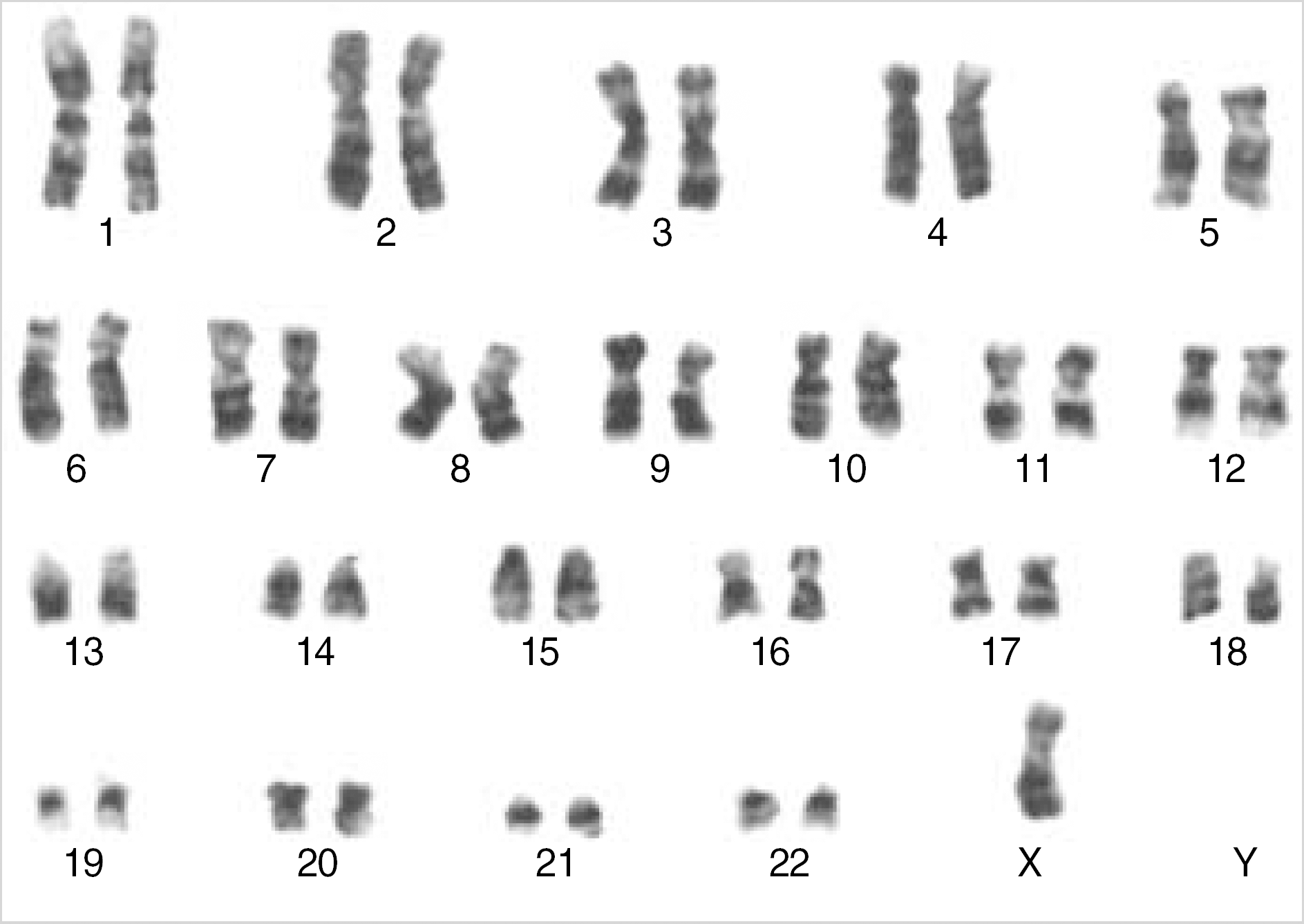
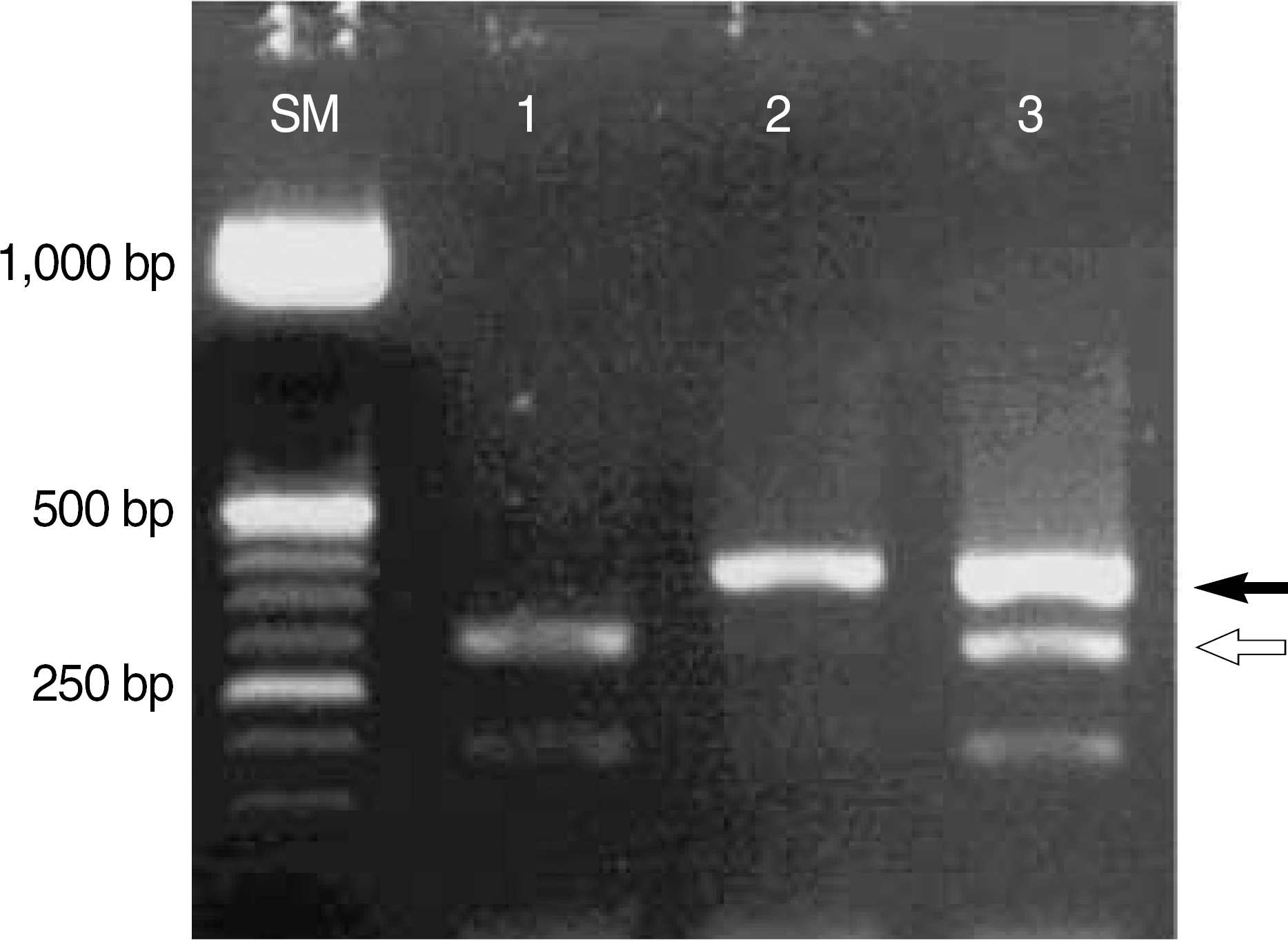
We report a case that revealed the characteristics of acute myeloblastic leukemia with maturation (AML-M2) on the morphology of the bone marrow biopsy and 45,X,-Y in conventional cytogenetic study, but was confirmed to have a typical AML1/ETO translocation by molecular studies using reverse transcriptase polymerase chain reaction and fluorescence in situ hybridization. Insertion of ETO gene on chromosome 8 into chromosome 21 in this patient resulted in the development of the chimeric gene, AML1/ETO, on the long arm of chromosome 21. Our final report on the patient's karyotype: 45,X,-Y.ish ins(21;8)(q22;q22q22)(AML1+,ETO+;ETO+,AML1-). In case typical morphologic features compatible with recurrent cytogenetic abnormalities are shown, molecular studies in addition to conventional cytogenetic study might be required to confirm the diagnosis.
References
1. Brunning RD, Matutes E. Acute myeloid leukemia with recurrent genetic abnormalities. Jaffe ES, Harris NL, editors. World Health Organization Classification of Tumours. Pathology and Genetics of Tumours of Haematopoietic and Lymphoid Tissues. Lyon: IARC press;2001. p. 81–3.
2. Bennett JM, Catovsky D, Daniel MT, Flandrin G, Galton DA, Gralnick HR, et al. Proposals for the classification of the acute leukaemias. French-American-British (FAB) co-operative group. Br J Haematol. 1976; 33:451–8.

3. Bennett JM, Catovsky D, Daniel MT, Flandrin G, Galton DA, Gralnick HR, et al. Proposed revised criteria for the classification of acute myeloid leukemia: A report of the French-American-British Cooperative Group. Ann Intern Med. 1985; 103:620–5.
4. Groupe Francias de Cytogenetique Hematologique. Acute myelogenous leukemia with an 8;21 translocation. A report on 148 cases. Cancer Genet Cytogenet. 1990; 44:169–79.
5. Saitoh K, Miura I, Ohshima A, Takahashi N, Kume M, Utsumi S, et al. Translocation (8;12;21)(q22.1;q24.1;q22.1): a new masked type of t(8;21)(q22;q22) in a patient with acute myeloid leukemia. Cancer Genet Cytogenet. 1997; 96:111–4.

6. Wong KF, Kwong YL, So CC. Translocation (8;20;21)(q22;q13;q22) in acute myeloblastic leukemia with maturation: a variant form of t(8;21). Cancer Genet Cytogenet. 1998; 101:39–41.
7. Harrison CJ, Radford-Weiss I, Ross F, Rack K, le Guyader G, Vekemans M, et al. Fluorescence in situ hybridization analysis of masked (8;21)(q22;q22) translocations. Cancer Genet Cytogenet. 1999; 112:15–20.

8. Gamerdinger U, Teigler-Schlegel A, Pils S, Bruch J, Viehmann S, Keller M, et al. Cryptic chromosomal aberrations leading to an AML1/ETO rearrangement are frequently caused by small insertions. Genes Chromosomes Cancer. 2003; 36:261–72.
9. Albano F, Specchia G, Anelli L, Liso A, Zagaria A, Santoro A, et al. Submicroscopic deletions in an acute myeloid leukemia case with a four-way t(8;11;16;21). Leuk Res. 2005; 29:855–8.

10. The Fourth International Workshop on Chromosomes in Leukemia: a prospective study of acute nonlymphocytic leukemia. Chicago, Illinois, USA, September 2–7, 1982. Cancer Genet Cytogenet. 1984; 11:284–7.
11. Berger R, Bernheim A, Daniel MT, Valensi F, Sigaux F, Flandrin G. Cytologic characterization and significance of normal karyotypes in t(8;21) acute myeloblastic leukemia. Blood. 1982; 59:171–8.

12. Gallego M, Carroll AJ, Gad GS, Pappo A, Head D, Behm F, et al. Variant t(8;21) rearrangements in acute myeloblastic leukemia of childhood. Cancer Genet Cytogenet. 1994; 75:139–44.

13. Andrieu V, Radford-Weiss I, Troussard X, Chane C, Valensi F, Guesnu M, et al. Molecular detection of t(8;21)/AML1-ETO in AML M1/M2: correlation with cytogenetics, morphology and immunopheno-type. Br J Haematol. 1996; 92:855–65.

14. Onozawa M, Fukuhara T, Nigo M, Takeda A, Takahata M, Yamamoto Y, et al. Insertion (21;8)(q22;q22q22): a masked t(8;21) in a patient with acute myelocytic leukemia. Cancer Genet Cytogenet. 2003; 147:134–9.

15. Sarriera JE, Albitar M, Estrov Z, Gidel C, Aboul-Nasr R, Manshouri T, et al. Comparison of outcome in acute myelogenous leukemia patients with translocation (8;21) found by standard cytogenetic analysis and patients with AML1/ETO fusion transcript found only by PCR testing. Leukemia. 2001; 15:57–61.

16. Miyagi J, Kakazu N, Masuda M, Miyagi T, Toyohama T, Nakazato T, et al. Acute myeloid leukemia (FAB-M2) with a masked type of t(8;21) translocation revealed by spectral karyotyping. Int J Hematol. 2002; 76:338–43.

17. Maseki N, Miyoshi H, Shimizu K, Homma C, Ohki M, Sakurai M, et al. The 8;21 chromosome translocation in acute myeloid leukemia is always detectable by molecular analysis using AML1. Blood. 1993; 81:1573–9.

18. Maruyama F, Yang P, Stass SA, Cork A, Freireich EJ, Lee MS, et al. Detection of the AML1/ETO fusion transcript in the t(8;21) masked translocation in acute myelogeneous leukemia. Cancer Res. 1993; 53:4449–51.
19. Maruyama F, Stass SA, Estey EH, Cork A, Hirano M, Ino T, et al. Detection of AML1/ETO fusion transcript as a tool for diagnosing t(8;21) positive acute myelogeneous leukemia. Leukemia. 1994; 8:40–5.
20. Langabeer SE, Walker H, Rogers JR, Burnett AK, Wheatley K, Swirsky D, et al. Incidence of AML1/ETO fusion transcripts in patients entered into the MRC AML trials. Br J Haematol. 1997; 99:925–8.
21. Lee DY, See CJ, Hwang CD, Cho HI, Lee DS. Analysis of discrepancies between G-banding and FISH in hematologic abnormalities. Korean J Clin Pathol. 2001; 21:445–50.
Fig. 1.
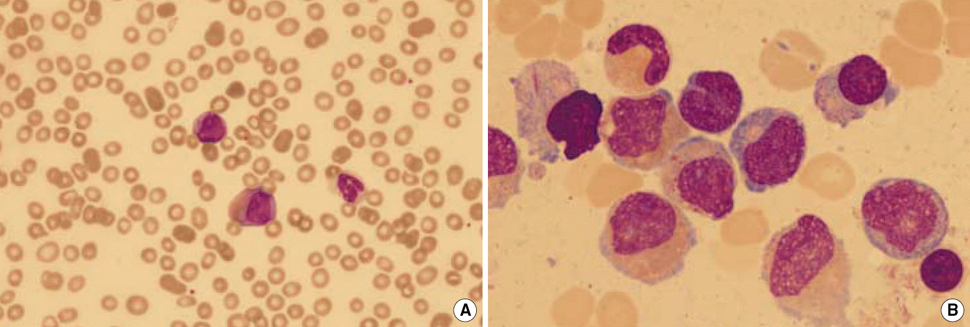
Leukemic cells in (A) peripheral blood smear (×400) and in (B) bone marrow aspirate smear (×1,000, Wight Giemsa stain). They reveal large sized, round to oval shaped nuclei with or without nucleoli and moderate to abundant cytoplasm with azurophilic granules. Some leukemic cells show large azurophilic granules (abnormal fusion), and Auer rods are occasionally found. They also show cytoplasmic staining abnormalities, including homogenous pink colored cytoplasm.

Fig. 3.
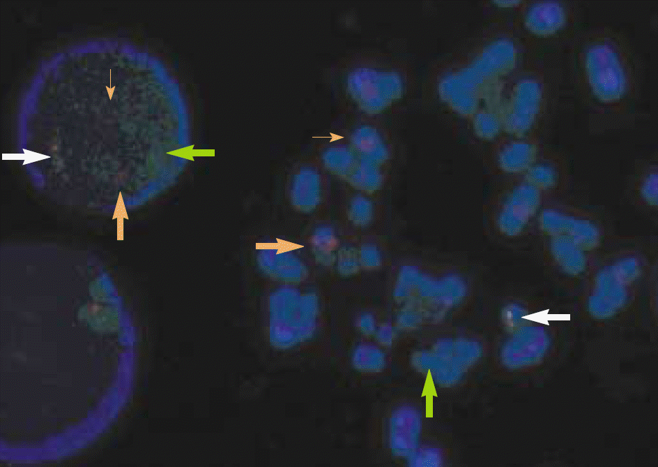
FISH analysis with LSI AML1 (orange)/ETO (green) dual color, dual fusion probe showed chromosome 8 (thick and thin orange arrows), one normal chromosome 21 (green arrow), and one fusion signal (white arrow) on chromosome 21, confirming the insertion of a fragment of ETO gene on chromosome 8 into the long arm of chromosome 21.


 PDF
PDF ePub
ePub Citation
 Print
Print


 XML Download