Abstract
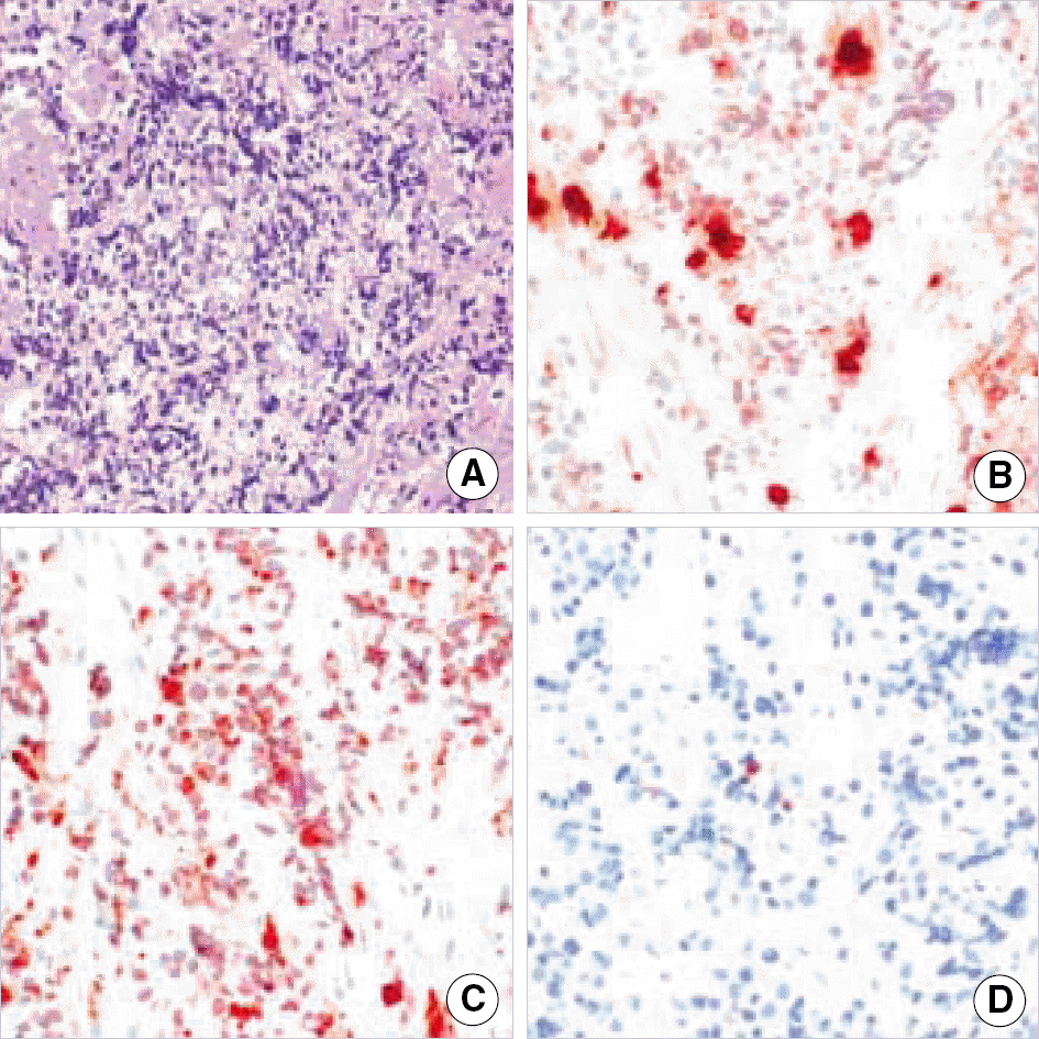
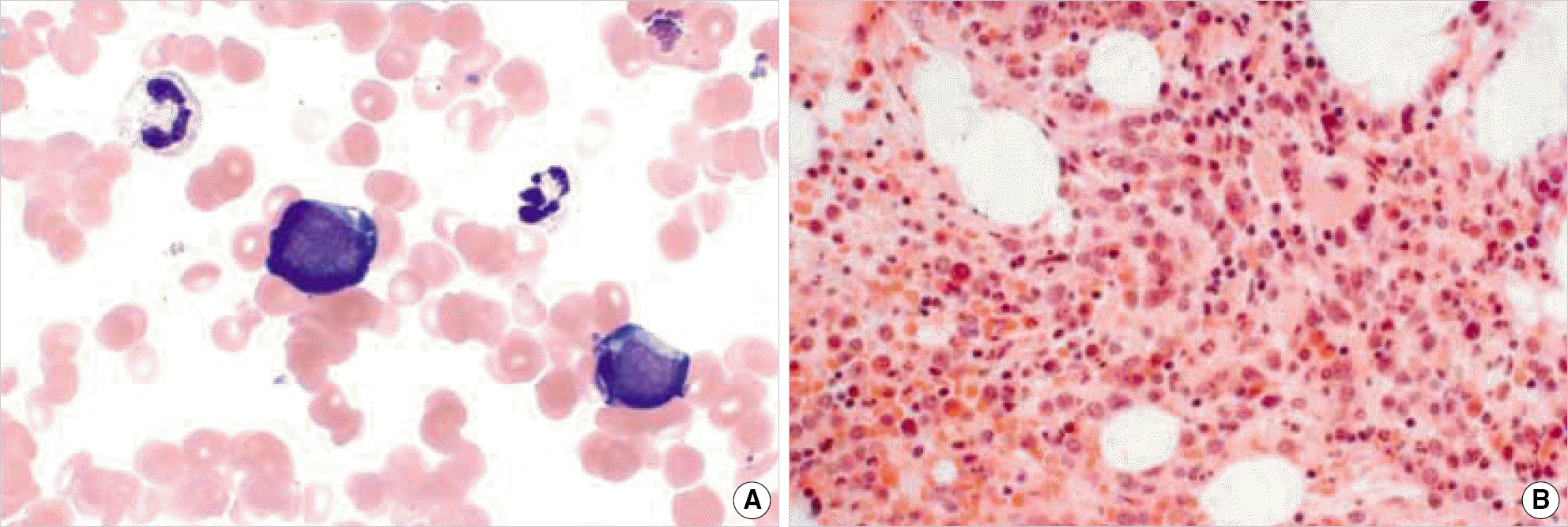
Granulocytic sarcoma of the uterine adnexa is a rare event. A 50-year-old woman, who had previously been diagnosed as chronic myeloid leukemia (CML), but had a complete hematologic response, presented with lower abdominal pain and a large pelvic mass involving the right uterine adnexa region and extending to the right posterior wall of the bladder and right distal ureter. A biopsy of the uterine adnexa revealed granulocytic sarcoma, and a subsequent bone marrow biopsy confirmed the diagnosis of CML in the blastic phase.
References
1. Delaflor-Weiss E, Zauber NP, Kintiroglou M, Berman EL, DeWitt R, Malcynski D. Acute myelogenous leukemia relapsing as granulocytic sarcoma of the cervix. A case Report. Acta Cytol. 1999; 43:1124–30.
2. Muss HB, Moloney WC. Chloroma and other myeloblastic tumours. Blood. 1973; 42:721–8.
3. Oliva E, Ferry JA, Young RH, Prat J, Srigley JR, Scully RE. Granulocytic sarcoma of the female genital tract: a clinicopathologic study of 11 cases. Am J Surg Pathol. 1997; 21:1156–65.

4. Pathak B, Bruchim I, Brisson ML, Hammouda W, Bloom C, Gotlieb WH. Granulocytic sarcoma presenting as tumors of the cervix. Gynecol Oncol. 2005; 98:493–7.

5. Neiman RS, Barcos M, Berard C, Bonner H, Mann R, Rydell RE, et al. Granulocytic sarcoma: a clinicopathologic study of 61 biopsied cases. Cancer. 1981; 48:1426–37.

6. Suh YK, Shin HJ. Fine-needle aspiration biopsy of granulocytic sarcoma: a clinicopathologic study of 27 cases. Cancer. 2000; 90:364–72.
7. Mwanda WO, Rajab JA. Granulocytic sarcoma: report of three cases. East Afr Med J. 1999; 76:594–6.
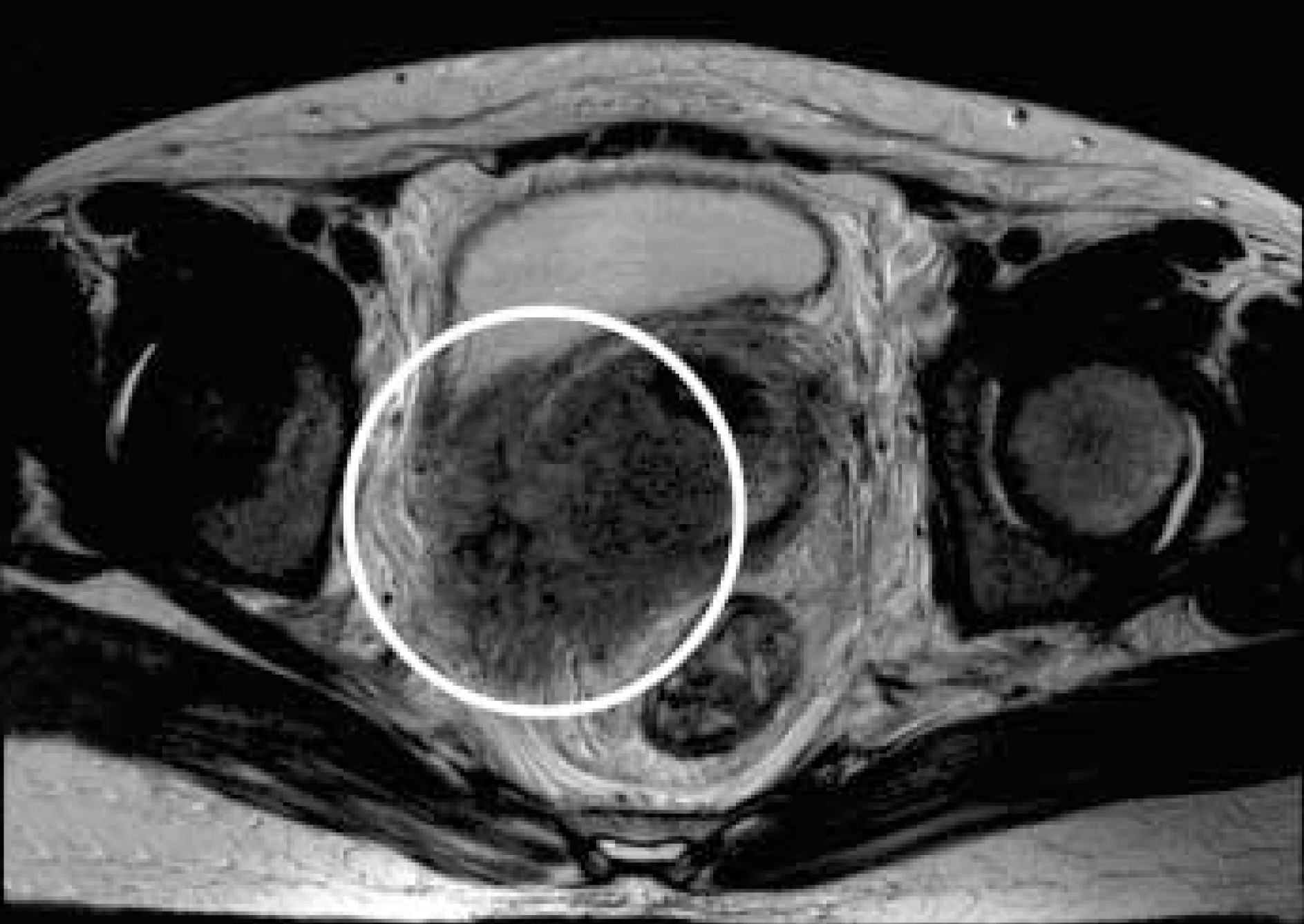
Fig. 1.
MRI of the pelvis with enhancement shows a mass of the right uterine adnexa region with a size of 6.5×6.0 cm infiltrating the wall of the bladder and uterus.


 PDF
PDF ePub
ePub Citation
 Print
Print


 XML Download