

 PDF
PDF ePub
ePub Citation
 Print
Print


INTRODUCTION
Extrapulmonary (EP) tuberculosis accounts for 5% of all case of tuberculosis. Anorectal tuberculosis is a rare extrapulmonary form of the disease. Diagnosis of tuberculous fistula depends mainly on suspicion of its clinical features such as multiple fistulas, recurrence of fistula, presence of concomitant pulmonary, intestinal tuberculosis and inguinal lymphadenopathy.(1) Also it is very difficult to distinguish tuberculous fistula from other granulomatous diseases such as Crohn's fistula. Traditionally, invasive procedures for microbiologic and pathologic diagnosis are usually required, and they are the diagnostic procedures of choice for tuberculous anal fistula. However, they have several limitations of availability, accuracy and reliability, and biopsy is of limited diagnostic value in the differentiation of tuberculous from Crohn's fistula.
The whole-blood interferon-gamma (IFN-γ) enzyme-linked immunosorbent assay (QuantiFERON-TB Gold in Tube test (QFT-GIT); Cellestis Limited, Victoria, Australia) has been used for the diagnosing of Mycobacterial tuberculosis infection including latent tuberculosis infection and tuberculosis disease.(2) This test was approved by the U.S. Food and Drug Administration (FDA) in 2007. Yet, its diagnostic performance for detection of tuberculous anal fistula has never been defined. The aim of this work was to study its diagnostic usefulness in individuals suspected of having tuberculous anal fistula.
METHODS
For the period of May 2007 to May 2009, we selected patients in whom a diagnosis of tuberculous anal fistula could be suspected on the basis of the following criteria: recurrent, multiple anal fistulae or abscesses, pulmonary, intestinal TB, or presence of epithelioid granulomas on histologic examination of the excised tissue and all participants underwent the GFT-GIT assay. The criteria for the diagnosis of tuberculous anal fistula was by 1) looking for Koch's bacillus in the anal lesion by direct examination (Ziehl-Neelsen stain), 2) presence of caseating epithelioid granulomas on histology, and 3) complete remission after anti-TB treatment.
1) QuantiFERON-TB gold
The QFT-GIT test was performed according to the manufacturer's instructions.(3) A total of 3 ml of peripheral venous blood was collected into 3 tubes of 1 ml each, which included a nil control tube, TB antigen tube, and an mitogen tube. The tubes were incubated within 16 hours of collection at 37℃ as soon as possible. Following a 16 to 24 hour incubation period, enzyme-linked immunosorbent assay (ELISA) for IFN-γ (IU/ml) was performed according to the manufacturer's instructions. A positive result was defined as TB antigen minus nil control IFN-γ when is greater than or equal to 0.35 IU/ml and increments of nil control IFN-γ levels to 25% or more.(3,4) An indeterminate result was defined as either a nil control IFN-γ level of >8.0 IU/ml or the value of the mitogen minus nil control of <0.5 IU/ml with a TB antigen minus a nil control IFN-γ level of either <0.35 IU/ml or <25% of the nil control value.
RESULTS
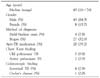
119 patients having suspected diagnoses of TB fistula were retrospectively enrolled. Of these, 51 patents (43%) were classified as having TB fistula, including 31 with the presence of chronic caseating granuloma and acid-fast bacilli (confirmed tuberculosis) and 20 with successful response to antituberculosis therapy (probable tuberculosis). The remaining 68 patients (57%) were classified as not having tuberculosis. Table 1 shows the clinical characteristics of the 51 patients with TB fistula. There were 43 males, and the median age was 43 (range, 16~76) years. In this study, 7 patients with TB fistula occurred co-existing with a pulmonary lesion and 4 patients with intestinal lesion.
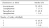
Fistulas were classified according to a modified Parks classification as intersphincteric (n=22), transsphincteric (n=7), suprasphincteric (n=21), and extrasphincteric (n=1). Nine patients had multiple fistulae (Table 2).
The diagnostic performance of the QFT-GIT assay are shown in Table 3. The sensitivity, specificity, PPV, NPV were 86%, 85%, 81%, and 89%, respectively.
DISCUSSION
According to the World Health Organization (2007), tuberculosis is spreading and currently a third of the world's population is infected with Mycobacterium tuberculosis.(5) Mycobacterial tuberculosis infection is still a public health problem both in underdeveloped and in developed countries where the epidemic of human immunodeficiency virus (HIV), the appearance of multiresistant bacilli, large immigrant populations and poverty all play their part in the increased incidence of the disease.(6) As such, tuberculosis can no longer be considered a rare disease in the United States.
Extrapulmonary tuberculosis can be found in any organ. Gastrointestinal TB comprises <1 percent of all cases and anoperineal disease is very rare. Anal fistula is the most frequent symptom of anorectal TB (80~91% of cases)(7-9) and anal TB is commonly seen in men (4:1 ratio) and in the 4th decade of life. The results concur with those of this study. Although most cases of TB fistula occur in the presence of active pulmonary disease,(10) only two cases occurred in the presence of active pulmonary tuberculosis and 5 cases had TB scar in chest radiographs without previous history of TB treatment in this study. Intestinal tuberculosis was also associated with TB fistula in 4 cases. The type of tuberculous fistula in ano is usually high and multiple fistulae in the majority and diagnosis of TB fistula depends mainly on suspicion of its clinical features.
As opposed to typical cryptoglandular anorectal fistula, tuberculous fistula can be cured only by medical treatment alone. Unfortunately, there is no functional sign or preferred site that allows a tuberculous fistula to be distinguished from a cryptoglandular fistula.(11) Although traditional diagnostic methods such as Ziehl-Neelsen stain (ZNS) and culture methods are available for suspected tuberculous fistula, ZNS test is characterized by poor sensitivity. About 40~60% of patients with pulmonary disease and 75% of patients with extrapulmonary disease go undiagnosed by this method. In this study, ZNS for M. Tuberculosis was positive only in 4 patients (7.8%) with dissatisfying results, as well. A minimum number of 104/ml bacilli are required for microscopy. Culture methods used in clinical laboratories are growth-dependent and may take 6~8 weeks to produce a negative/positive result. Recently, polymerase chain reaction (PCR) has been reported for detection of a specific sequence for M. tuberculosis directly in clinical specimens.(12) But there were only limited data involving a small number (only 4) of patients.(13)
The typical histologic features of TB lesion is caseating eithelioid cell granuloma, but the pathognomic presence of specific caseation is not constant and presents diagnostic problems. So, it is difficult to differentiate tuberculous from Crohn's fistula because of similar clinical, pathological, radiological, and endoscopic findings. Granulomatous findings from biopsied specimens are a common feature in TB and CD. Other possible granulomatous diseases of the anus can occur, such as amoebiasis, in reaction to a foreign body, sarcoidosis, syphillis, and venereal lymphogranuloma, etc.(14,15) Although attempts have been made to distinguish them, there are still no specific differential diagnostic methods up until now.
Interferon-gamma assay was assessed as a potential candidate to replace the Mantoux skin test and was approved by the U.S. FDA in 2005. The QFT-G is an ELISA test that measures the release of interferon gamma in blood from sensitized persons. The antigens consist of synthetic peptides representing 2 M. tuberculosis proteins, early secretory antigenic target 6 (ESAT-6) and culture filtrate protein 10 (CFP10). Blood is incubated with the antigens, and interferon gamma released by sensitized leukocytes is measured.(16,17) Its advantages include that it necessitates only a single patient visit, results are available in 24 hours, and the findings are not affected by prior BCG vaccination. In 2007, the FDA approved the next generation Interferon-gamma release assay, the QuantiFERON-TB Gold in Tube test (QFT-GIT). This test contains an extra antigen, TB7.7, which theoretically improves sensitivity and circumvents the time-consuming step of manually stimulating lymphocytes, as the tubes already contain the antigens.(17,18)
Recently there have been several reports on GFT-GIT for extrapulmonary tuberculosis, and results showed sensitivities for diagnosis of EP-TB ranging from 14% to 80%.(19,20) However, In our study, QFT-GIT assay showed 86% sensitivity and 85% specificity for diagnosis of tuberculous anal fistula. These results revealed a high diagnostic accuracy compared to other studies.

 XML Download