

 PDF
PDF ePub
ePub Citation
 Print
Print


INTRODUCTION
Accurate prediction of lymph node (LN) status is of crucial importance for appropriate curative treatment planning in early gastric cancer (EGC). EGCs without LN metastasis can be curatively treated with minimally invasive endoscopic resection such as endoscopic mucosal resection or endoscopic submucosal dissection, whereas LN positive EGCs should be referred to gastrectomy with limited or extended LN dissection.(1) Compared to surgical treatment, selected cases of LN negative EGCs treated by endoscopic resection show excellent prognosis, in addition to minimal morbidity and mortality and better postoperative quality of life.(2) Therefore, the aggressive surgical approach should be reserved only for EGC patients at risk of LN metastasis.
In an attempt for proper prediction of LN status in EGC, several studies have identified the pathologic characteristics that are associated with an increased likelihood of LN metastasis,(3) which are currently used as eligibility criteria for endoscopic resection. Recently, with an advancement of molecular biology in gastric cancer, some molecular markers, such as, the PCNA labeling index,(4) p53 expression,(5) erbB-2 expression,(6) DNA ploidy,(7) and VEGF expression,(8) are also emerging as novel markers for predicting LN status in EGC.
Matrix metalloproteinases (MMPs) are a family of enzymes that are responsible for the breakdown of connective tissue proteins in the extracellular matrix. It plays central roles in the tissue remodeling associated with growth, development, and tissue repair under normal physiologic condition.(9) Recently, it has been demonstrated that aberrant MMPs expressions contribute to the pathogenesis of metastasis and tumor progression in several human malignancies. More specifically, the following MMPs have been reported to be involved in the tumor progression and metastasis of gastric carcinoma; membrane- type MMP (MT1-MMP),(10) MMP-1,(11) MMP-2,(12,13) MMP-3,(14) MMP-7,(15,16) and MMP-9.(17) In this study, to investigate the association between MMPs expressions and LN metastasis in EGC, we immunohistochemically analyzed MMPs expressions in the tumor samples of pT1 gastric cancers.
METHODS
1) Patients
Between May 2004 and April 2006, there were 512 EGC patients treated with gastrectomy and regional LN dissection at Chonnam National University Hwasun Hospital. Of these, 34 (6.6%) cases had LN metastasis at final pathologic examinations and were included in this study. Eighty EGC patients without LN metastasis were collected at random and used as controls. All patients underwent standard gastrectomy including distal or total gastrectomy with limited or extended LN dissection. Gastric cancer samples were obtained with informed consent from the Chonnam National University Hwasun.
2) Histopathology
After resection, all specimens were fixed in 10% buffered formalin, embedded in paraffin wax, and serially sectioned at 3~5 mm. Sections were stained with hematoxylin and eosin (H&E) and subjected to pathological examination, at which depth of invasion, lymph node metastasis, histologic type, Lauren's classification, and lymphovascular permeation (as outlined by the Japanese Classification of Gastric Carcinoma) were determined.(18) Degrees of histologic differentiation and tumor stages were determined as detailed in the 6th edition of the TNM Classification System of the UICC.(19) Sections containing greatest tumor areas or the invasive front were selected, and each section was serially sectioned at 4µm for immunohistochemistry (IHC).
3) Immunohistochemistry
Tissue sections from formalin-fixed, paraffin-embedded blocks were deparaffinized with xylene, rehydrated through graded ethanol, rinsed with distilled water, and washed with Tris-buffered saline (TBS). Antigen retrieval was performed by boiling for 20 min in citrate buffer (pH 6.0). Avidin-biotin-peroxidase complex staining using DAB as a chromogen was performed using a standard technique, as described by the manufacturer (Dako, Glostrup, Denmark). The primary antibodies used were rabbit anti-MMP-2 (1:100, Santa Cruz Biotechnology, Santa Cruz, CA), rabbit anti-MMP-9 (1:100, Santa Cruz Biotechnology, Santa Cruz, CA), mouse anti-MMP-7 (1:75, Chemicon), mouse anti-CK (1:100, Dako, Glostrup, Denmark), and mouse anti-D2-40 (1:50, Dako, Glostrup, Denmark).
4) Evaluation of immunostaining
Staining for MMP-2, MMP-7, and MMP-9 at the invasive front were evaluated by two independent researchers (JH Lee and YK Park) without knowledge of clinicopathological data, and staining intensities (stains were dark brown in color) were graded as:0, negative; 1+, weak staining; 2+, moderate; and 3+, strong. MMP immunostaining was deemed positive when moderate to strong membranous and/or cytoplasmic staining was observed at the invasive front (the percentages of positively stained tumor cells were not counted). Cytokeratin staining was used to improve the visualization of small numbers of tumor cells budding from invasive edges. Tumor budding was classified into four grades; none, minimal, moderate, or severe, as previously described.(20) Degree of tumor budding was then categorized as low-grade (none or minimal) and high-grade (moderate to severe). D2-40 staining was used to differentiate lymphatic vessels and the endothelial cells of blood vessels.
5) Statistical analysis
SPSS version 12.0 (SPSS, Chicago, IL) was used throughout. Categorical comparisons of the clinicopathological characteristics of the study groups were carried out using the chi-square or Fisher's exact test. To determine the significances of associations between variables and nodal metastasis, matched data were subjected to conditional logistic regression analysis. For all statistical tests, P-values of less than 0.05 were considered to be statistically significant.
RESULTS
1) Clinicopathological characteristics associated with lymph node metastasis
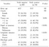
The study cohort consisted of 73 men and 41 women of mean age 60 years. No differences were found between the LN positive and LN negative group with respect to age or gender. Pathological examination revealed significant inter-group differences with respect to tumor size, depth of invasion, and lymphatic invasion between LN positive and LN negative group (Table 1). Tumor size was significantly larger in the LN positive group (P=0.001), and there were significantly more submucosal invasion (82.4% vs. 35.5%, P<0.001) and lymphatic invasion (64.7% vs. 10.0%, P<0.001) in the LN positive group.
2) Immunohistochemistry
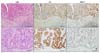
Immunohistochemistry showed MMP-7 expression in 28 (82.4%) of the 34 LN positive pT1 tumor samples, which was significantly higher than that in the LN negative group (82.4% vs. 54.4%, P=0.005) (Table 2). MMP-7 expression was observed in the cytoplasm and cell membranes of tumor cells, especially in deeper part of the tumor regions, namely at the invasive front (Fig. 1). MMP-9 expression was also greater in the LN positive group, but with marginal statistical significance (85.3% vs. 67.5%, P=0.051). However, no significant inter-group difference was found for MMP-2 expression (70.6% in LN positive group vs. 57.5% in LN negative group, P=0.189).
3) Predicting factors for lymph node metastasis
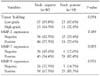
To identify predicting factors for LN metastasis in pT1 tumors, variables found to be significantly associated with LN metastasis in histopathological examinations and IHC, such as, tumor size, submucosal invasion, lymphatic invasion, and MMP-7 expression, were subjected to univariate and multivariate analysis (Table 3). Multivariate analysis was performed using a logistic regression model which incorporated all of these variables, and revealed that MMP-7 expression (OR 4.915, 95% CI 1.375~17.573) and the presences of lymphatic invasion (OR 10.337, 95% CI 2.785~38.360) were independent predictors for LN metastasis in pT1 tumors.
DISCUSSION
Increasing evidence indicates that the extracellular matrix (ECM) of tumors and non-cancerous stromal cells play an important role in tumor progression and metastasis.(21) Multistep phenomena involving the proteoloytic degradations of basement membranes and ECM reduce cellular adhesion and increase the ability of tumor cells to relocate, and these processes are known to be prerequisites of tumor cell invasion and metastasis.(22) Matrix metalloproteinases (MMPs) are a subfamily of the metzincin proteases, which is one of several families of metalloendopeptidases. Mechanisms of ECM protein degradation by MMPs regulate various cellular characteristics, such as, cell growth, differentiation, apoptosis, and migration under physiologic condition.(9) Recently, it has been demonstrated that the expressions and activities of MMPs are upregulated in virtually all human cancers.(23)
Minimally invasive endoscopic resection, such as endoscopic mucosal resection or endoscopic submucosal dissection, is a treatment of choice for LN negative EGCs in Asian regions.(1) Compared to surgical treatment, it offers several advantages including minimal morbidity and mortality and better postoperative quality of life as well as excellent long term outcomes when performed in selected patients.(24) Given the increasing use of endoscopic treatment for EGC, it is critical that we are able to determine the LN status of EGC for appropriate selection of patients for this treatment. Current indications used for predicting LN status in EGCs are generally based on the histopathological parameters that suggest the high likelihood of the LN metastasis. EGCs of ulcerative type, undifferentiated histology, lymphovascular invasion, or submucosal invasion are candidates for surgical treatment due to the high risk of LN metastasis.(25) However, established eligibility criteria for endoscopic resection for EGC are debated, and there is no definitive consensus yet on which patients and/or tumor characteristics are associated with LN metastasis in EGC.(3,26)
Recently, with a better understanding of molecular biology in gastric cancer, several molecular-pathological markers have been investigated to determine their associations with LN metastasis in EGC. PCNA labeling index,(4) p53 expression,(5) erbB-2 expression,(6) DNA ploidy,(7) and VEGF expression,(8) are good examples that have been shown to be associated with LN metastasis in EGC. In this study, although sample size is small, we found that MMP-7 expression is also associated with LN metastasis in EGC. In our analysis, MMP-7 expression remained as an independent predicting factor for LN metastasis even after adjusting other pathologic parameters. To our best knowledge, this study is the first to demonstrate the relation of MMP-7 expression to LN metastasis in EGC.
MMP-7 has broad-spectrum proteolytic activity against a variety of ECM substrates, including type IV collagens, proteoglycans, laminin, fibronectin, and casein.(9) Unlike other MMPs, the expression of MMP-7 is predominantly restricted to carcinoma cells in deeply invading tumor cell nests.(16) Yamashita and colleagues(27) showed that its expression levels are directly correlated with vessel invasion and metastasis in human gastric cancer. Aihira and colleagues(28) also showed an association between MMP-7 expression and submucosal invasion and lymph node metastasis in early stage signet ring cell gastric carcinoma. Similarly, the carcinoma cell-specific expression of MMP-7 and its association with tumor invasiveness and prognosis have also been demonstrated in other malignancies such as pancreatic,(29) esophageal,(30) and colorectal carcinoma.(15) Their findings are consistent with our result indicating that MMP-7 may play an important role in the early stages of tumor invasion and LN metastasis in human gastric cancer.
In the present study, MMP-2 and MMP-9 were found to be more highly expressed in pT1 tumors with LN metastasis, but did not reach statistical significance. In the literature, MMP-2 is one of the most extensively investigated MMPs, and has been shown to be significantly associated with tumor invasion, vascular permeation, and lymph node metastasis in gastric cancer.(13) In addition, a significant correlation between MMP-2 expression and prognosis has been suggested.(12) MMP-9 also has been shown to be associated with tumor invasiveness and survival in gastric cancer, and its expression has also been associated with initial gastric cancer invasion.(17) Therefore, considering small sample size of our study, we think we need more studies with larger patients to investigate the association of MMP-2 and MMP-9 with LN metastasis in EGC.
Summarizing, the present study shows that MMP-7 expression is associated with LN metastasis in EGC, which suggests that MMP-7 may have an important role during the early stages of LN metastasis in gastric cancer. Our results suggest that MMP-7 expression can be used as a molecular-pathologic marker for predicting LN metastasis in EGC.

 XML Download