

 PDF
PDF ePub
ePub Citation
 Print
Print


INTRODUCTION
Early gastric cancer (EGC) is defined as being confined to the mucosa or the submucosa, regardless of regional lymph-node metastasis.(1) If it has metastatic nodes, the curability depends on the dissection of metastatic lymph nodes. But, it is hard to evaluate metastatic lymph nodes by either endoscopic ultrasound (EUS) or computed tomography (CT).(2,3) Recently, endoscopic mucosal resection (EMR)/endoscopic submucosal dissection (ESD) has been widely accepted for standard and useful treatment of early gastric cancer as it is less invasive, conserving the whole stomach and improving postoperative quality of life.(4-7) Although several attempts have been made to predict metastatic lymph nodes for early gastric cancer, there have been some reports evaluating criteria for additional surgical treatment.(8-13) In general, both EMR and ESD were performed on the following conditions. 1) differentiated (well- and/or moderately differentiated adenocarcinoma and/or papillary carcinoma) type, mucosal cancer without ulcer, and any size, 2) differentiated type, mucosal cancer with ulcer, and 3 cm or smaller, 3) undifferentiated type, mucosal cancer without ulcer, and 2 cm or smaller, 4) no lymphovascular invasion and 5) depth of tumor' invasion, confined to the submucosal 1 (SM1; ~500µm).(1,14,15)
Recently, in our institution, we extended indications of ESD for patients with mucosal cancer without ulcer findings irrespective of tumor size, mucosal cancer with ulcer findings (≤3 cm) and minute (<500 m from muscularis mucosae) submucosal invasive cancer (≤2 cm). All patients with poorly differentiated type adenocarcinoma were excluded from ESD.
In our institution, before the ESD, EUS (Endoscopic Ultra-Sonography) and CT (Computed Tomography) gastrography were performed to evaluate the stage of patients.
In the present study, we therefore introduced the necessity of surgical treatment after EMR/ESD of EGC.
METHODS
1) Patient population and specimens
Between April 2005 and December 2009, 140 consecutive patients had received additional gastrectomy with lymph node dissection after the procedures of EMR/ESD in hospital. All EMR/ESD were performed by specialized enterologists. A single-channel endoscope (GIF-Q260, Olympus, Tokyo, Japan) was uses in patients under conscious sedation. Lesions were marked beyond a 5 mm margin using a conventional needle knife. A solution of epinephrine and methylene blue in normal saline was injected into the submucosal layer. Submucosal dissection with circumferential incision was performed using an insulated-tip diathermic (IT) knife. To confirm resection margin, the specimens containing tumors were sliced at intervals of 2 mm. In EMR procedures, the only difference was that dissection was performed by direct snaring using an oval device.
In this study, the criteria for surgical treatment were 1) mucosal cancer, with undifferentiated-type histology (>2 cm) or involvement of margin and 2) submucosal cancer. In all patients, the extent of lymph node dissection was over D1+β. The surgically removed lymph nodes were histologically examined for metastasis.
2) Statistical analysis
Statistical analysis was performed using the "Statistical Package for Social Science (SPSS)," version 12.0 for Windows (SPSS, Inc, Chicago, IL). In analysis of metastatic lymph nodes, we excluded 3 patients who developed advanced gastric cancer after surgery. All parameters, considered tobe candidatesforrisk factorsin metastatic lymph nodes, were investigated by univariate and multivariate analyses. The χ2 test was used for univariate analysis. Ap value ofless than 0.05 was considered as significant. Multivariate analysis was done with variables significant in univariate analysis using binary logistic multiple regression tests with backward elimination.
RESULTS
1) Demographics of patients
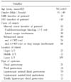
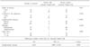
The clinical characteristics of 140 patients are presented in Table 1. The ESD was performed in 130 of the 140 patients. In mucosal lesion, 50 patients had additional surgery for undifferentiated-type histology (>2 cm) and lateral margin involvement. 105 of the 140 patients showed lesions at the lower part of the stomach. Laparoscopic gastrectomy was performed in 93 patients.
2) Incidence of residual cancer after EMR/ESD
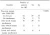
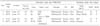
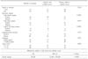
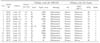
In the pathological results, 29 of the 66 patients with incomplete resection margin showed residual cancer after gastrectomy. Thirty-six patients with only lateral margin involvement showed high residual cancer compared with vertical margin. Also, patients who were diagnosed with incomplete vertical resection margin showed advanced gastric cancer in the final pathologic results. Especially, 3 of the 74 patients with clear resection margin showed residual cancer after surgical treatment (Table 2). 3 of the 4 patients with deep submucosal invasion (Depth >1,000µm or very close deep resection margin or deep margin involvement) developed advanced gastric cancer after surgery (Table 3). In univariate analysis for residual cancer, lateral margin, lateral and deep margin, and large tumor were significant predictive factors. Only lateral margin showed statistical significance in multivariate analysis (Table 4).
3) Incidence of lymph node metastasis
In univariate analysis, submucosal invasion with advanced depth (over sm2 or 500µm) and the presence of lymphovascular invasion had the statistical significance for metastatic lymph nodes. But in multivariate analysis, only the presence of lymphovascular invasion showed predictive factors for lymph node metastasis (Table 5).
The details of patients with lymph node metastasis are described in Table 6. One patient with mucosal cancer had a metastatic lymph node. He had the longest diameter of 5.3 cm and the presence of lymphovascular invasion. Two patients with lymph node metastasis were in the sm1 group and had lymphovascular invasion.
DISCUSSION
EGC has high disease-specific five-year survival rate (over 95%) after surgical treatment.(16-20) EMR/ESD is the standard and preferred treatment for many patients with EGC without the risk of lymph node metastasis.(4,5) Also, ESD makes it possible to resect large lesions en bloc, permitting precise pathological results and decreasing the recurrence of cancer.(21) But after EMR/ESD, additional surgical treatment is also needed to remove residual cancer or metastatic lymph nodes in some cases. Surgical treatment was determined by the pathological results of specimens obtained from EMR/ESD. Resection margin involvement including lateral or deep margin was an important indication of additional surgical treatment to eliminate residual cancer.(22,23) The results of this study showed that the patients with lateral margin involvement needed additional surgical resection to remove residual cancer. But, although there was no significance statistically for deep margin involvement, additional surgical resection should be performed to eliminate the possibility of residual cancer. We speculate that the low ratio of residual cancer concerning deep margin involvement resulted from the singeing effects of the IT Knife during dissection. Also, deep invasion of submucosal lesion (over sm2 or 500µm) also was an important indication for surgery to remove metastatic lymph nodes.(8,15,22) Some of the deep submucosal invasion with clear resection margin showed residual cancer or more advanced gastric cancer in the final pathological results. And, the presence of lymphovascular invasion was an important predictive factor for metastatic lymph nodes.
As stated above, the patients with lateral margin involvement showed high residual cancer rates. On the other hand, patients with deep margin involvement showed low residual cancer rates in the final pathologic results. But unlike lateral margin, one patient with deep resection margin involvement displayed advanced gastric cancer after gastrectomy. Besides this, 3 patients with clear resection margin showed residual cancer or more advanced gastric cancer. Especially, 2 of the 3 patients displayed advanced gastric cancer after gastrectomy. In terms of tumor invasion, it is conceivable that deep submucosal invasion (deep margin involvement or over sm2 group) had the possibility of more advanced gastric cancer compared with the tumor depth of ESD. Because the diathermic knife could provide a clear resection margin from the tumor, additional gastrectomy inevitable requires confirmation for more advanced gastric cancer in patients with deep submucosal invasion.
In the present study, 13 of 140 patients displayed metastatic lymph nodes after gastrectomy. One patient with lymph node metastasis displayed advanced gastric cancer (depth of invasion, subserosa layer). Excepting 3 patients who were diagnosed with advanced gastric cancer after surgery, deepsubmucosal invasion (over sm2 or 500µm) and the presence of lymphovascular invasion showed significant differences for metastatic lymph nodes. Especially in multivariate analysis, the presence of lymphovascular invasion was a predictive factor for metastatic lymph nodes. Therefore additional surgical treatment was positively necessary for patient with lymphovasular invasion or deepsubmucosal invasion.
In conclusion, because EMR/ESD of EGC is less traumatic than surgery, EMR/ESD should be considered as a first-line treatment in selected patients without risk of residual cancer or lymph node metastasis. But, additional surgical management should be done to eliminate the possibility of residual cancer or more advanced gastric cancer or metastatic lymph nodes for patients with deepsubmucosal invasion (over sm2 or 500µm) or presence of lymphovascular invasion.
 XML Download