

 PDF
PDF ePub
ePub Citation
 Print
Print


Abstract
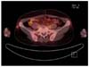
MFH is rare with just a few thousand cases diagnosed each year. We report a case of right flank malignant fibrous histiocytoma (MFH) in the short-term after tumor excision. A 47-year-old woman visited hospital after being diagnosed with MFH. Resection margin was positive, so we planned for wide excision. While awaiting surgery, 1.5 cm sized new mass occurred adjacent to the incision site. The patient underwent wide excision. Postoperative pathologic findings observed a 1.0 cm diameter mass with infiltrative borders in the subcutaneous fat. It was composed of spindle or epithelial cell showing severe polymorphism. Many mitotic cells were observed including atypia. In immunohistochemical study, tumor cells were negative for smooth muscle actin, desmin, myoglobin, S100, cytokeratin, and positive for CD68, and thus diagnosed as pleomorphic MFH. With the purpose of improving local tumor control, radiation therapy was performed after wide excision.
Figures and Tables
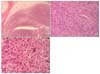 | Fig. 2(A) Microscopic findings of the right flank mass. The tumor has un encapsulated ill-defined margin infiltrating surrounding subcutaneous adipose tissue (H&E, ×12.5). (B) Microscopic findings of the right flank mass. The spindle tumor cells grow in fascicular pattern (H&E, ×100). (C) Microscopic findings of the right flank mass. The tumor cells have markedly pleomorphic nuclei with frequent mitoses (H&E, ×400).
|
References
1. Weiss SW, Goldblum JR. Soft Tissue Tumors. 2008. 5th ed. Philadelphia: Mosby.
2. Fletcher CDM, Unni KK, Mertens F. WHO Classification of Tumors, Pathology & Genetics Tumors of Soft Tissue and Bone. 2002. Lyon: IARC Press.
3. Le Doussal V, Coindre JM, Leroux A, Hacene K, Terrier P, Bui NB, et al. Prognostic factors for patients with localized primary malignant fibrous histiocytoma. Cancer. 1996. 77:1823–1830.
4. Salo JC, Lewis JJ, Woodruff JM, Leung DH, Brennan MF. Malignant fibrous histiocytoma of the extremity. Cancer. 1999. 85:1765–1772.
5. Weiss SW, Enzinger FM. Malignant fibrous histiocytoma. An analysis of 200 cases. Cancer. 1978. 41:2250–2256.
6. Yang JC, Chang AE, Baker AR, Sindelar WF, Danforth DN, Topalian SL, et al. Randomized prospective study of the benefit of adjuvant radiation therapy in the treatment of soft tissue sarcomas of the extremity. J Clin Oncol. 1998. 16:197–203.
7. Coindre JM, Terrier P, Bui NB, Bonichon F, Collin F, Le Doussal V, et al. A study of 546 patients from the French Federation of Cancer Centers Sarcoma Group. Prognostic factors in adult patients with locally controlled soft tissue sarcoma. J Clin Oncol. 1996. 14:869–877.
8. Gaynor JJ, Tan CC, Casper ES, Collin CF, Friedrich C, Shiu M, et al. Refinement of clinicopathologic staging for localized soft tissue sarcoma of the extremity. J Clin Oncol. 1992. 10:1317–1329.
9. Pisters PW, Leung DH, Woodruff J, Shi W, Brennan MF. Analysis of prognostic factors in 1041 patients with localized soft tissue sarcomas of the extremities. J Clin Oncol. 1996. 14:1679–1689.
 XML Download