

 PDF
PDF ePub
ePub Citation
 Print
Print


Abstract
Purpose
Isolated superior mesenteric artery (SMA) dissection is a rare, but increasing vascular disorder. However, optimal treatment guidelines are not well established. The purpose of this study is to review a single institutional experience in the management of isolated SMA dissections and establish optimal treatment guidelines.
Methods
Between November 2004 and August 2009, 26 patients were diagnosed with isolated SMA dissection at Eulji University Hospital. Diagnosis was confirmed with CT scans in all patients. We retrospectively reviewed the medical records, imaging studies, and the early outcomes of the patients.
Results
There were 22 (84.5%) men and 4 women. The mean age was 55.4 (39~74) years. The mean follow-up was 39.1 (4.1~53.3) months. In 15 patients, CT scans were performed for abdominal pain, and in the other 11 patients, the isolated SMA dissections were detected incidentally during workup for other causes. The radiographic findings included an intimal flap with a patent false lumen in 16 and intramural hematoma in 10. The dissection started at a mean of 22.3 (5~46) mm from the origin of the SMA with a mean length was 47.7 (10~150) mm. Treatments included expectant management in 13, anticoagulation in 6, stenting in 6 patients, and surgery in one case of bowel infarction. None required additional intervention. All patients remained asymptomatic during follow-up.
Figures and Tables
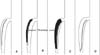 | Fig. 1Classification of CT findings shows double lumen without stenosis (A), intramural thrombosis without stenosis (B), intramural thrombosis with stenosis (C), double lumen with closed false lumen and stenosis (D).
|
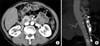 | Fig. 2Incidental finding. (A) SMA has intramural thrombosis without stenosis. (B) Double lumen with intimal flap begins from proximal SMA.
|
References
1. Gobble RM, Brill ER, Rockman CB, Hecht EM, Lamparello PJ, Jacobowitz GR, et al. Endovascular treatment of spontaneous dissections of the superior mesenteric artery. J Vasc Surg. 2009. 50:1326–1332.
2. Morris JT, Guerriero J, Sage JG, Mansour MA. Three isolated superior mesenteric artery dissections: update of previous case reports, diagnostics, and treatment options. J Vasc Surg. 2008. 47:649–653.
3. Subhas G, Gupta A, Nawalany M, Oppat WF. Spontaneous isolated superior mesenteric artery dissection: a case report and literature review with management algorithm. Ann Vasc Surg. 2009. 23:788–798.
4. Sakamoto I, Ogawa Y, Sueyoshi E, Fukui K, Murakami T, Uetani M. Imaging appearances and management of isolated spontaneous dissection of the superior mesenteric artery. Eur J Radiol. 2007. 64:103–110.
5. Okada M, Ishiguchi T, Itoh H. Management of spontaneous dissection of the superior mesenteric artery. Intern Med. 2004. 43:451–452.
6. Suzuki S, Furui S, Kohtake H, Sakamoto T, Yamasaki M, Furukawa A, et al. Isolated dissection of the superior mesenteric artery: CT findings in six cases. Abdom Imaging. 2004. 29:153–157.
7. Yasuhara H, Shigematsu H, Muto T. Self-limited spontaneous dissection of the main trunk of the superior mesenteric artery. J Vasc Surg. 1998. 27:776–779.
8. Nakamura K, Nozue M, Sakakibara Y, Kuramoto K, Satoh M, Kobayashi S, et al. Natural history of a spontaneous dissecting aneurysm of the proximal superior mesenteric artery: report of a case. Surg Today. 1997. 27:272–274.
9. Kim HK, Kwon TW, Cho YP, Kim GE. Treatment of an isolated superior mesenteric artery dissection. J Korean Soc Vasc Surg. 2007. 23:159–162.
10. Ambo T, Noguchi Y, Iwasaki H, Kondo J, Matsumoto A, Suzuki H, et al. An isolated dissecting aneurysm of the superior mesenteric artery: report of a case. Surg Today. 1994. 24:933–936.
11. Nagai T, Torishima R, Uchida A, Nakashima H, Takahashi K, Okawara H, et al. Spontaneous dissection of the superior mesenteric artery in four cases treated with anticoagulation therapy. Intern Med. 2004. 43:473–478.
12. Sisteron A, Vieville C. Jausseran JM, Reggi M, Courbier R, editors. Observations personnelles. Chirurgie des Arteriopathies Digestives. 1975. Paris: Expansion Scientifique Francaise;197–202.
13. Picquet J, Abilez O, Penard J, Jousset Y, Rousselet MC, Enon B. Superficial femoral artery transposition repair for isolated superior mesenteric artery dissection. J Vasc Surg. 2005. 42:788–791.
14. Leung DA, Schneider E, Kubik-Huch R, Marincek B, Pfammatter T. Acute mesenteric ischemia caused by spontaneous isolated dissection of the superior mesenteric artery: treatment by percutaneous stent placement. Eur Radiol. 2000. 10:1916–1919.
15. Froment P, Alerci M, Vandoni RE, Bogen M, Gertsch P, Galeazzi G. Stenting of a spontaneous dissection of the superior mesenteric artery: a new therapeutic approach? Cardiovasc Intervent Radiol. 2004. 27:529–532.
16. Kim JH, Roh BS, Lee YH, Choi SS, So BJ. Isolated spontaneous dissection of the superior mesenteric artery: percutaneous stent placement in two patients. Korean J Radiol. 2004. 5:134–138.
17. Casella IB, Bosch MA, Sousa WO Jr. Isolated spontaneous dissection of the superior mesenteric artery treated by percutaneous stent placement: case report. J Vasc Surg. 2008. 47:197–200.
 XML Download