

 PDF
PDF ePub
ePub Citation
 Print
Print


Abstract
A solitary fibrous tumor (STF) is a relatively unusual neoplasm first described as a distinctive tumor arising from pleura. Some reports have shown that STF also affect extrathoracic regions. A 70-year-old woman was referred to our hospital for surgical treatment of an incidentally discovered thigh mass. We performed complete removal of the tumor. It was a soft tissue tumor with muscle indentation but without invasion to the surrounding muscles. The resected specimen was 7.0×6.3×5.2 cm. Histologically, the tumor was composed of a haphazard proliferation of spindle cells and epitheloid cells with hypercellularity and high mitotic activity. Immunohistochemistry showed positive immunoreactivity for CD34, CD99, bcl-2 protein, CD117, vimentin, smooth muscle actin and epithelial membrane antigen. We report, herein, on a rare case of malignant SFT in the thigh region along with a review of the literature.
Figures and Tables
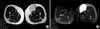 | Fig. 2MRI images of the mass in the thigh. A MRI shows 6.6×5.5×4.7 cm sized well-defined lobulated mass in the subcutaneous fat layer of left thigh. (A) Homogeneous low SI on T1WI, (B) heterogeneous high SI on T2WI.
|
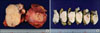 | Fig. 3Gross findings of the thigh mass. (A) The mass shows 7.0×6.3×5.2 cm sized, oval shaped, soft and friable tumor and cut surfaces are homogenously cream yellow to pinkish purple with a fish-flesh appearance. (B) The cut surfaces are multifocally spotty petechially hemorrhagic and focal minimally necrotic.
|
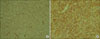
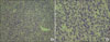 | Fig. 4Malignant solitary fibrous tumor. (A) The tumor shows hemangiopericytomatous pattern of mainly spindle & partly epitheloid tumor cells (H&E stain, ×200). (B) Mainly spindle & partly epitheloid tumor cells have relatively high mitotic activity and focal mild nuclear atypia & pleomorphism (H&E stain, ×400).
|
References
1. Klemperer P, Rabin CB. Primary neoplasms of the pleura: a report of five cases. Arch Pathol. 1931. 11:385–412.
2. Akisue T, Matsumoto K, Kizaki T, Fujita I, Yamamoto T, Yoshiya S, et al. Solitary fibrous tumor in the extremity: case report and review of the literature. Clin Orthop Relat Res. 2003. (411):236–244.
3. Moran CA, Suster S, Koss MN. The spectrum of histologic growth patterns in benign and malignant fibrous tumors of the pleura. Semin Diagn Pathol. 1992. 9:169–180.
4. Gold JS, Antonescu CR, Hajdu C, Ferrone CR, Hussain M, Lewis JJ, et al. Clinicopathologic correlates of solitary fibrous tumors. Cancer. 2002. 94:1057–1068.
5. Park MS, Araujo DM. New insights into the hemangiopericytoma/solitary fibrous tumor spectrum of tumors. Curr Opin Oncol. 2009. 21:327–331.
6. Guillou L, Fletcher JA, Fletcher CDM, Mandahl N. Fletcher CDM, Unni KK, Mertens F, editors. Extrapleural solitary fibrous tumor and hemangiopericytoma. Pathology and Genetics of Tumours of Soft Tissue and Bone. 2002. Lyon: IARC Press;86–90.
7. Si Y, Kim HJ, Kang WK, Jung CK, Oh ST. Malignant solitary fibrous tumor in the perianal region. J Korean Surg Soc. 2007. 73:443–446.
8. Anders JO, Aurich M, Lang T, Wagner A. Solitary fibrous tumor in the thigh: review of the literature. J Cancer Res Clin Oncol. 2006. 132:69–75.
9. Lee SC, Tzao C, Ou SM, Hsu HH, Yu CP, Cheng YL. Solitary fibrous tumors of the pleura: clinical, radiological, surgical and pathological evaluation. Eur J Surg Oncol. 2005. 31:84–87.
10. Gengler C, Guillou L. Solitary fibrous tumour and haemangiopericytoma: evolution of a concept. Histopathology. 2006. 48:63–74.
 XML Download