

 PDF
PDF ePub
ePub Citation
 Print
Print


Abstract
Purpose
Pneumatosis intestinalis (PI) is increasingly being detected in recent years with the more frequent use of computerized tomography (CT). The present study was performed to evaluate the clinico-radiologic characteristic presentation of PI and to determine the prognostic factors for mortality.
Methods
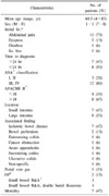
Fifteen patients who were diagnosed with PI on CT between June 2000 and May 2010 were retrospectively reviewed. Age, sex, location of PI, presence of portal vein gas, time to diagnosis, American Society of Anesthesiologists (ASA) classification, Acute Physiology And Chronic Health Evaluation II (APACHE II), acidosis, shock, and other associated findings were analyzed for their association with outcome.
Results
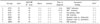
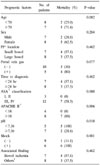
Fifteen patients (7 males and 8 females, average age, 60.3 years) were diagnosed with PI. Mortality rate was 47% (7 patients). The mortality rate in patients with septic shock, APACHE II score (18), acidosis (pH<7.36) were all 100%, 87%, 100%, respectively (P<0.05). Age, sex, location of PI, portal vein gas, time to diagnosis, ASA classification, associated findings did not show statistical difference.
Figures and Tables
Fig. 1
CT finding of pneumatosis intestinalis. (A) Contrast enhanced CT scan shows linear gas collection involving long segment of distal ileum. (B) Lung window setting shows clearly gas in intramural space.
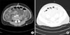
Fig. 2
Operative finding of pneumatosis intestinalis. Small bowel is edematous and has necrotic change on distal ileum segment.
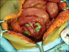
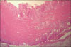
Fig. 3
Pathologic finding of pneumatosis intestinalis. Bowel wall shows transmural hemorrhagic infarction (H&E stain, ×10).
References
1. DuVernoi JC. Anatomische beobachtungen der unter der aeusseren und inneren haut der gedaerme eingeschlossenen. Luft Phys Med Abhandl Acad Wissensch Petersburg. 1783. 2:182.
2. Goodman RA, Riley TR 3rd. Lactulose-induced pneumatosis intestinalis and pneumoperitoneum. Dig Dis Sci. 2001. 46:2549–2553.
3. Heng Y, Schuffler MD, Haggitt RC, Rohrmann CA. Pneumatosis intestinalis: a review. Am J Gastroenterol. 1995. 90:1747–1758.
4. Morris MS, Gee AC, Cho SD, Limbaugh K, Underwood S, Ham B, et al. Management and outcome of pneumatosis intestinalis. Am J Surg. 2008. 195:679–682.
5. Kim HL, Lee HK, Park SJ, Yi BH, Ko BM, Hong HS, et al. Pneumatosis intestinalis: CT findings and clinical features. J Korean Radiol Soc. 2008. 58:149–154.
6. Wolters U, Wolf T, Stützer H, Schröder T. ASA classification and perioperative variables as predictors of postoperative outcome. Br J Anaesth. 1996. 77:217–222.
7. Chung HH, Seo YJ, Choi JS, Kim JH. Significance of APACHE score in patients with a gastrointestinal perforation. J Korean Surg Soc. 1998. 55:809–817.
8. Koss LG. Abdominal gas cysts (pneumatosis cystoides intestinorum hominis); an analysis with a report of a case and a critical review of the literature. AMA Arch Pathol. 1952. 53:523–549.
9. Chippindale AJ, Desai S. Two unusual cases of pneumatosis coli. Clin Radiol. 1991. 43:180–182.
10. Jamart J. Pneumatosis cystoides intestinalis. A statistical study of 919 cases. Acta Hepatogastroenterol (Stuttg). 1979. 26:419–422.
11. Nelson SW. Extraluminal gas collections due to diseases of the gastrointestinal tract. Am J Roentgenol Radium Ther Nucl Med. 1972. 115:225–248.
12. Yale CE, Balish E. Pneumatosis cystoides intestinalis. Dis Colon Rectum. 1976. 19:107–111.
13. Caudill JL, Rose BS. The role of computed tomography in the evaluation of pneumatosis intestinalis. J Clin Gastroenterol. 1987. 9:223–226.
14. Kelvin FM, Korobkin M, Rauch RF, Rice RP, Silverman PM. Computed tomography of pneumatosis intestinalis. J Comput Assist Tomogr. 1984. 8:276–280.
15. Ham JH, Kim TH, Han SW, Cho KJ, Choi SO, Pack JS, et al. A case of pneumatosis cystoides intestinalis: diagnosed by CT colonoscopy. Korean J Gastroenterol. 2007. 50:334–339.
16. Park YS. A case of pneumatosis cystoides intestinalis. J Korean Surg Soc. 1976. 18:55–62.
17. Togawa S, Yamami N, Nakayama H, Shibayama M, Mano Y. Evaluation of HBO2 therapy in pneumatosis cystoides intestinalis. Undersea Hyperb Med. 2004. 31:387–393.
18. Elliott GB, Elliott KA. The roentgenologic pathology of so called pneumatosis cystoides intestinalis. Am J Roentgenol Radium Ther Nucl Med. 1963. 89:720–729.
19. Wiesner W, Mortelé KJ, Glickman JN, Ji H, Ros PR. Pneumatosis intestinalis and portomesenteric venous gas in intestinal ischemia: correlation of CT findings with severity of ischemia and clinical outcome. AJR Am J Roentgenol. 2001. 177:1319–1323.
20. Greenstein AJ, Nguyen SQ, Berlin A, Corona J, Lee J, Wong E, et al. Pneumatosis intestinalis in adults: management, surgical indications, and risk factors for mortality. J Gastrointest Surg. 2007. 11:1268–1274.
 XML Download