

 PDF
PDF ePub
ePub Citation
 Print
Print


Abstract
Purpose
The aim of this study was to evaluate the usefulness of sentinel lymph node (SLN) biopsy in the treatment of primary melanoma.
Methods
Fifty-one cases that were diagnosed as malignant melanoma of the skin without clinical evidence of regional lymph node metastasis and underwent SLN biopsy at Samsung Medical Center were analyzed retrospectively. A lymphoscintigraphy with peritumoral injection of radionuclide was performed preoperatively. SLNs were identified using a hand-held gamma probe and by methylene blue dye injection intraoperatively.
Results
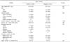
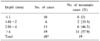
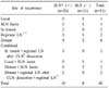
Twenty patients (39%) had metastasis in the SLN and they underwent immediate complete radical dissection of the nodal basin. Among the 20 patients who had SLN metastasis, additional metastatic lymph nodes were detected in 5 patients after the complete lymph node dissection. When several clinico-pathologic parameters such as gender, age, primary tumor location, draining nodal basin, tumor depth and size of tumor were compared between SLN positive group and negative group, there was a significant difference in the mean thickness of melanoma between SLN (+) group (5±2.9 mm) and SLN (-) group (4.5±5.0 mm) (P<0.05). In the same way, as the thickness of melanoma increased, positive SLN were detected more frequently (P<0.05). Recurrences occurred in 18 patients (35.3%) during the follow-up period, but only one case in 31 patients with negative SLN recurred at the SLN basin without evidence of distant or loco-regional recurrence (false negative rate 4.8%). Lymphedema of extremity developed in 9 patients who underwent complete radical lymph node dissection and 2 patients who underwent only SLN biopsy had a very mild-form lymphedema.
Figures and Tables
Fig. 1
F-18 fluorodeoxyglucose (FDG) positron emission tomography. It shows a malignant lesion on right finger tip (arrow).
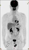
Fig. 2
Tc-99m labeled lymphoscintigraphy. Sentinel lymph node is identified as a hot uptake in right axillary area.
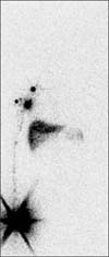
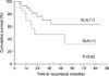
Fig. 3
Recurrence free survival between sentinel lymph node (SLN) positive group and SLN negative group.
References
1. Jemal A, Siegel R, Ward E, Hao Y, Xu J, Murray T, et al. Cancer statistics, 2008. CA Cancer J Clin. 2008. 58:71–96.
2. Community of Population-based Regional Cancer Registries in Korea. An estimation of the national cancer incidence in Korea for 2000~2002 using the databases of 8 population-based regional cancer registries. J Prev Med Public Health. 2008. 41:380–386.
3. Gershenwald JE, Thompson W, Mansfield PF, Lee JE, Colome MI, Tseng CH, et al. Multi-institutional melanoma lymphatic mapping experience: the prognostic value of sentinel lymph node status in 612 stage I or II melanoma patients. J Clin Oncol. 1999. 17:976–983.
4. Balch CM, Soong SJ, Gershenwald JE, Thompson JF, Reintgen DS, Cascinelli N, et al. Prognostic factors analysis of 17,600 melanoma patients: validation of the American Joint Committee on Cancer melanoma staging system. J Clin Oncol. 2001. 19:3622–3634.
5. Sim FH, Taylor WF, Pritchard DJ, Soule EH. Lymphadenectomy in the management of stage I malignant melanoma: a prospective randomized study. Mayo Clin Proc. 1986. 61:697–705.
6. Coates AS, Ingvar CI, Petersen-Schaefer K, Shaw HM, Milton GW, O'Brien CJ, et al. Elective lymph node dissection in patients with primary melanoma of the trunk and limbs treated at the Sydney Melanoma unit from 1960 to 1991. J Am Coll Surg. 1995. 180:402–409.
7. Morton DL, Wen DR, Wong JH, Economou JS, Cagle LA, Storm FK, et al. Technical details of intraoperative lymphatic mapping for early stage melanoma. Arch Surg. 1992. 127:392–399.
8. Alex JC, Weaver DL, Fairbank JT, Rankin BS, Krag DN. Gamma-probe-guided lymph node localization in malignant melanoma. Surg Oncol. 1993. 2:303–308.
9. Gershenwald JE, Tseng CH, Thompson W, Mansfield PF, Lee JE, Bouvet M, et al. Improved sentinel lymph node localization in patients with primary melanoma with the use of radiolabeled colloid. Surgery. 1998. 124:203–210.
10. Rousseau DL Jr, Ross MI, Johnson MM, Prieto VG, Lee JE, Mansfield PF, et al. Revised American Joint Committee on Cancer staging criteria accurately predict sentinel lymph node positivity in clinically node-negative melanoma patients. Ann Surg Oncol. 2003. 10:569–574.
11. Lee TH, Shim JS, Jeong JH. Lymphoscintigraphy for intraopertive sentinel node biopsy of skin and soft tissue malignancy. J Korean Soc Plast Reconstr Surg. 2005. 32:635–640.
12. Kim HS, Song SK, Sim SJ, Kang DY, Kim KH. Sentinel lymph node biopsy and staging of melanoma using lymphoscintigraphy and gamma-probe. Korean J Dermatol. 2003. 41:1575–1582.
13. Oh DS, Roh RT, Yoo WM, Park C, Park BY. Management of malignant melanoma using sentinel lymph node biopsy: a case report. J Korean Soc Plast Reconstr Surg. 2003. 30:651–654.
14. Kim CW, Huh D, Lee CJ. Treatment of malignant melanoma using sentinel lymph node dissection. Korean J Dermatol. 2003. 41:58–64.
15. McMasters KM, Reintgen DS, Ross MI, Gershenwald JE, Edwards MJ, Sober A, et al. Sentinel lymph node biopsy for melanoma: controversy despite widespread agreement. J Clin Oncol. 2001. 19:2851–2855.
16. Doting MH, Hoekstra HJ, Plukker JT, Piers DA, Jager PL, Tiebosch AT, et al. Is sentinel node biopsy beneficial in melanoma patients? A report on 200 patients with cutaneous melanoma. Eur J Surg Oncol. 2002. 28:673–678.
17. Rutkowski P, Nowecki ZI, Nasierowska-Guttmejer A, Ruka W. Lymph node status and survival in cutaneous malignant melanoma--sentinel lymph node biopsy impact. Eur J Surg Oncol. 2003. 29:611–618.
18. Thompson JF, McCarthy WH, Bosch CM, O'Brien CJ, Quinn MJ, Paramaesvaran S, et al. Sentinel lymph node status as an indicator of the presence of metastatic melanoma in regional lymph nodes. Melanoma Res. 1995. 5:255–260.
19. Nowecki ZI, Rutkowski P, Nasierowska-Guttmejer A, Ruka W. Sentinel lymph node biopsy in melanoma patients with clinically negative regional lymph nodes--one institution's experience. Melanoma Res. 2003. 13:35–43.
20. Seok JW, Kim IJ. Camparison of the efficiency for Tc-99m Tin-colloid and Tc-99m phytate in sentinel node detection in breast cancer patients. Nucl Med Mol Imaging. 2008. 42:451–455.
21. Vaggelli L, Castagnoli A, Borgognoni L, Urso C, Matteini M, Cesco P. Radioisotopic lymphatic mapping of the sentinel node in melanoma: importance of immunohistochemistry. Tumori. 2000. 86:346–348.
22. Li LX, Scolyer RA, Ka VS, McKinnon JG, Shaw HM, McCarthy SW, et al. Pathologic review of negative sentinel lymph nodes in melanoma patients with regional recurrence: a clinicopathologic study of 1152 patients undergoing sentinel lymph node biopsy. Am J Surg Pathol. 2003. 27:1197–1202.
23. Houghton AN, Coit DG, Daud A, Dilawari RA, Dimaio D, Gollob JA, et al. Melanoma. J Natl Compr Canc Netw. 2006. 4:666–684.
24. Cuéllar FA, Vilalta A, Rull R, Vidal-Sicart S, Palou J, Ventura PJ, et al. Small cell melanoma and ulceration as predictors of positive sentinel lymph node in malignant melanoma patients. Melanoma Res. 2004. 14:277–282.
25. Niakosari F, Kahn HJ, McCready D, Ghazarian D, Rotstein LE, Marks A, et al. Lymphatic invasion identified by monoclonal antibody D2-40, younger age, and ulceration: predictors of sentinel lymph node involvement in primary cutaneous melanoma. Arch Dermatol. 2008. 144:462–467.
26. Carlson GW, Page AJ, Cohen C, Parker D, Yaar R, Li A, et al. Regional recurrence after negative sentinel lymph node biopsy for melanoma. Ann Surg. 2008. 248:378–386.
27. Yee VS, Thompson JF, McKinnon JG, Scolyer RA, Li LX, McCarthy WH, et al. Outcome in 846 cutaneous melanoma patients from a single center after a negative sentinel node biopsy. Ann Surg Oncol. 2005. 12:429–439.
28. Morton DL, Thompson JF, Cochran AJ, Mozzillo N, Elashoff R, Essner R, et al. Sentinel-node biopsy or nodal observation in melanoma. N Engl J Med. 2006. 355:1307–1317.
29. Chao C, Wong SL, Ross MI, Reintgen DS, Noyes RD, Cerrito PB, et al. Patterns of early recurrence after sentinel lymph node biopsy for melanoma. Am J Surg. 2002. 184:520–524.
30. Koskivuo I, Talve L, Vihinen P, Mäki M, Vahlberg T, Suominen E. Sentinel lymph node biopsy in cutaneous melanoma: a case-control study. Ann Surg Oncol. 2007. 14:3566–3574.
 XML Download