

 PDF
PDF ePub
ePub Citation
 Print
Print


INTRODUCTION
Pilomatrixoma is a benign, circumscribed, calcifying, epithelial neoplasm that arises from hair follicle matrix cells first described in 1880.(1)
Pilomatrixoma is usually found in young individuals and rarely presents in older people. It commonly occurs on the scalp, face, and neck, but is also found with lower frequency on the back or extremities. Pilomatrixoma usually presents as a solitary tumor, with multifocal types being very rare. It appears as a firm to hard, non-painful, ovalshaped tumor that is covered by normal skin.(2)
The aim of this study was to review our experience in regard to clinical symptoms, treatment, and outcomes of pilomatrixomas in a single institution, and to compare the clinicopathological features according to their location.
METHODS
We retrospectively reviewed the medical records of patients treated for pilomatrixoma at Gangnam Severance Hospital, Yonsei University College of Medicine, Seoul, Korea between January 1986 and December 2007.
Patients were divided into two groups according to their location of the lesion; patients with pilomatrixoma located on the head and neck were classified as Group I, and on the body as Group II.
We compared and analyzed between two groups for the demographic characteristics including sex distribution, age at initial presentation, age at operation, clinical manifestation, tumor size, histological finding, and treatment outcome. The mean follow-up period was 95.6 months (range, 9~207 months).
RESULTS
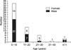
We identified 57 patients with a total 61 pilomatrixomas (four patients presented with multiple occurrences). The mean age at operation was 12.4 years (range, 1~68 years), and the majority of patients were in their first (59.7%), second (22.8%), and third (10.5%) decades of life. The sex distribution was approximately 1:1 (30 males, 27 females), but there was a predilection to males in the first decade and a predilection to females in the third decade of life (Fig. 1). Twenty-six tumors (42.6%) were located on the head, 16 (26.2%) on the neck, 15 (24.6%) on the extremities, and 4 (6.6%) on the trunk. Four patients presented with multiple occurrences, either concurrently (n=3, face and arm; scalp and shoulder; and eyelid and periauricular) or at a new site following treatment of the first lesion (n=1, back and arm). The most common initial presentation was a palpable mass (91.8%), and two patients also presented with pain and inflammation. The mean duration of symptoms was 20.4 months (range, 1~120 months), with most lasting less than one year. The mean tumor diameter was 1.46 cm (range, 0.4~4.0 cm), and the majority of tumors were below 2.0 cm (88.5%). All tumors were removed surgically. One patient with a pilomatrixoma in the parotid gland underwent superficial parotidectomy. Two patients relapsed, five months and three years later, and they were treated by surgical intervention repeatedly.
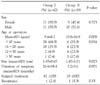
According to the tumor location, 42 tumors (68.9%) on the head and neck were classified as Group I, and 19 tumors (31.1%) on the body were classified as Group II.
The demographic features between two groups were listed in Table 1. Sex distribution was similar between two groups, and tumor size was also similar; 1.45 cm in Group I and 1.47 cm in Group II (P=0.023). The mean age at operation was 9.4 years in Group I, and 19.8 years in Group II with a significant statistical difference (P=0.009), and the mean duration of symptoms was 26.4 months in Group I, and 7.2 months in Group II (P=0.001).
DISCUSSION
Pilomatrixomas are tumors of ectodermal origin that arise from the outer root sheath cell of the hair follicle.(3) Although originally named "calcifying epitheliomas",(1) they were renamed "pilomatrixoma" after further histochemical and electron microscopic examination showed that these tumors arose from the outer root sheath cells of the hair follicle.(3)
Pilomatrixomas are most frequently found in the head and neck region, but they may occur anywhere, including the extremities and trunk. To date, there have been no reports of pilomatrixomas on the palms, soles, or genital region.(4) In our series, head and neck was the most common involved site (42.6%).
Pilomatrixomas usually occur within the first 2 years of life, 60% prior to age 10 years,(5-7) although they may also appear in adults. Pilomatrixomas usually show male predominance.
In our series, we also found that most of patients are less than 30 years (82.5%), a finding that is similar to previously accepted. However, our data showed no male predominance. The sex distribution was approximately 1:1 (30 males, 27 females), and there was a predilection to males in the first decade and a predilection to females in the third decade of life.
Comparing the demographic features of Groups I and II, the mean age at diagnosis in Group II was older than Group I with a significant statistical difference (19.8 years versus 9.4 years, P=0.009); however, the mean duration of symptoms was longer in Group I than Group II (26.4 months versus 7.2 months, P=0.001).
These results are thought to be that the pilomatrixomas located on the head and neck were found in early ages, but had a tendency to undergo the treatment unhurriedly.
Pilomatrixomas frequently present as single nodules,(8) but multifocal occurrences have been reported.(9,10) Multiple pilomatrixomas have been usually associated with disorders such as Gardner syndrome, Steinert disease, and sarcoidosis.(10) Tumor diameter varies from 0.5 cm to 15.0 cm, with most tumors being between 0.5 cm and 3.0 cm.(7) In our series, mean primary tumor diameter was 1.46 cm, and four patients (7.0%) had multifocal lesions not associated with above-mentioned disorders.
The most common presenting symptom is a hard, slowgrowing, and subcutaneous mass,(11) which is also shown in our study. Pilomatrixomas are characterized by calcification within the lesions, leading to a hard and bony feel and often resulting in an angulated shape (the 'tent' sign). These tumors are usually asymptomatic, but some have been associated with pain during episodes of inflammation or ulceration. Diagnosis of pilomatrixoma can be made histologically and clinically if the characteristics of the tumor are known. Radiologic imaging is of little diagnostic value. Plain radiograph show nonspecific calcification,(12) and computed tomography and magnetic resonance imaging only add detail to the surrounding structures and depth to the lesion. Fine needle aspiration cytology may reveal the presence of ghost cells, basaloid cells, and/or calcium deposition in the mass, all of which are diagnostic of pilomatrixoma.(13)
In histological findings, pilomatrixoma is a subcutaneous tumor occurring between the dermis and hypodermis, with medial displacement of the pilosebaceous glands and follicles. The most common microscopic features are islands of well-organized Malpighian cells; maturation is progressive and regular, with no atypical cells. The cells in the islands are arranged circularly, with nucleated basophilic cells on the periphery and enucleated shadow cells in the center. The islands are associated with a foreign body-type macrophagic reaction and fibrosis. In most cases, the lesions are poorly delineated, but encapsulated forms have also been observed (Fig. 2).(8,12)
Malignant forms are rare and characterized by a larger epithelial cell component, clusters of undifferentiated basaloid cells, the presence of atypical cells, invasion of blood vessels, and infiltration of capsular tissue.(14)
Since spontaneous regression is never observed, complete surgical excision is the treatment of choice and standard therapy for benign pilomatrixoma. Wide resection margins are recommended to minimize the risk of local recurrence.(5,7) Although overlying skin will need to be excised secondary to tumor adherence to the dermis occasionally, the tumor is rarely adherent to subcutaneous tissue and separation from underlying tissue is always easy. Recurrences after complete excision are rare, and the risk of local recurrence decreases progressively with age.(14,15)
In our series, two patients had a local recurrence after 5 months and 3 years respectively, but they were well controlled by re-excision completely.
In conclusion, pilomatrixoma is more common in children than in adults. It is a benign lesion and adequate local excision is the treatment of choice. Correct management of the patient can be maximized by awareness of the condition, careful clinical examination, and correct proper surgical operation.

 XML Download