

 PDF
PDF ePub
ePub Citation
 Print
Print


Abstract
Purpose
Papillary Thyroid Microcarcinoma (PTMC) is rapidly increasing due to increased interests in the public health care system and improvements in ultrasonographic instruments and fine-needle-aspiration technique. The aim of this study is to investigate relationships between clinicopathologic features and molecular markers of PTMC and to help in developing therapeutic strategies in PTMC.
Methods
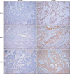
Tissue samples from patients with 38 PTMC and 21 benign thyroid tumors that were operated on from Jan. 2006 to Nov. 2008 were used to make microarrays and immunohistochemical staining for ER-α, E-CD, VEGF, MMP-2, MMP-9, and HIF-1α were performed. Clinicopathologic features of each immunohistochemical staining group were analyzed retrospectively.
Results
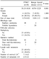
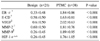
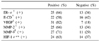
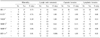
There is no immunohistochemistry staining in cases with benign thyroid lesions. The expression rate of ER-α, E-CD, VEGF, MMP-2, MMP-9, and HIF-1α in PTMC group was 66%, 58%, 82%, 66%, 71% and 63%, respectively. Bilateral tumor was statistically significant (48.0% vs 7.7%, P=0.015) related to MMP-2(+) PTMC group than in MMP-2(-) group. Bilateral tumor (44.4% vs 9.1%, P=0.060) and lymphovascular invasion (25.9% vs 0%, P=0.084) seemed to have greater relation to MMP-9(+) PTMC group than to MMP-9(-) group, but there is no statistically significant difference. Bilateral tumor (50.0% vs 7.1%, P=0.012), lymph node metastasis (45.8% vs 0%, P=0.003) and lymphovascular invasion (29.2% vs 0%, P=0.033) were significantly related to HIF-1α (+) PTMC group compared to HIF-1α(-) group.
Figures and Tables
References
1. Davies L, Welch HG. Increasing incidence of thyroid cancer in the United States, 1973-2002. JAMA. 2006. 295:2164–2167.
2. Ministry of Health and Welfare. Annual Report of the Korea Central Cancer Registry 2002. 2007.
3. Sherman SI. Thyroid carcinoma. Lancet. 2003. 361:501–511.
4. Hodgson NC, Button J, Solorzano CC. Thyroid cancer: is the incidence still increasing? Ann Surg Oncol. 2004. 11:1093–1097.
5. Choi YJ, Park YL, Koh JH. Prevalence of thyroid cancer at a medical screening center: pathological features of screen-detected thyroid carcinomas. Yonsei Med J. 2008. 49:748–756.
6. Beasley NJ, Lee J, Eski S, Walfish P, Witterick I, Freeman JL. Impact of nodal metastases on prognosis in patients with well-differentiated thyroid cancer. Arch Otolaryngol Head Neck Surg. 2002. 128:825–828.
7. Kilicarslan AB, Ogus M, Arici C, Pestereli HE, Cakir M, Karpuzoglu G. Clinical importance of vascular endothelial growth factor (VEGF) for papillary thyroid carcinomas. APMIS. 2003. 111:439–443.
8. Scarpino S, Cancellario d'Alena F, Di Napoli A, Pasquini A, Marzullo A, Ruco LP. Increased expression of Met protein is associated with up-regulation of hypoxia inducible factor-1 (HIF-1) in tumour cells in papillary carcinoma of the thyroid. J Pathol. 2004. 202:352–358.
9. von Wasielewski R, Rhein A, Werner M, Scheumann GF, Dralle H, Potter E, et al. Immunohistochemical detection of E-cadherin in differentiated thyroid carcinomas correlates with clinical outcome. Cancer Res. 1997. 57:2501–2507.
10. Klein M, Picard E, Vignaud JM, Marie B, Bresler L, Toussaint B, et al. Vascular endothelial growth factor gene and protein: strong expression in thyroiditis and thyroid carcinoma. J Endocrinol. 1999. 161:41–49.
11. Hay ID, Grant CS, van Heerden JA, Goellner JR, Ebersold JR, Bergstralh EJ. Papillary thyroid microcarcinoma: a study of 535 cases observed in a 50-year period. Surgery. 1992. 112:1139–1146. discussion 46-7.
12. Baudin E, Travagli JP, Ropers J, Mancusi F, Bruno-Bossio G, Caillou B, et al. Microcarcinoma of the thyroid gland: the Gustave-Roussy Institute experience. Cancer. 1998. 83:553–559.
13. Manole D, Schildknecht B, Gosnell B, Adams E, Derwahl M. Estrogen promotes growth of human thyroid tumor cells by different molecular mechanisms. J Clin Endocrinol Metab. 2001. 86:1072–1077.
14. Lee ML, Chen GG, Vlantis AC, Tse GM, Leung BC, van Hasselt CA. Induction of thyroid papillary carcinoma cell proliferation by estrogen is associated with an altered expression of Bcl-xL. Cancer J. 2005. 11:113–121.
15. Zeng Q, Chen GG, Vlantis AC, van Hasselt CA. Oestrogen mediates the growth of human thyroid carcinoma cells via an oestrogen receptor-ERK pathway. Cell Prolif. 2007. 40:921–935.
16. Cho MA, Lee MK, Nam KH, Chung WY, Park CS, Lee JH, et al. Expression and role of estrogen receptor alpha and beta in medullary thyroid carcinoma: different roles in cancer growth and apoptosis. J Endocrinol. 2007. 195:255–263.
17. Yane K, Kitahori Y, Konishi N, Okaichi K, Ohnishi T, Miyahara H, et al. Expression of the estrogen receptor in human thyroid neoplasms. Cancer Lett. 1994. 84:59–66.
18. Egawa C, Miyoshi Y, Iwao K, Shiba E, Noguchi S. Quantitative analysis of estrogen receptor-alpha and -beta messenger RNA expression in normal and malignant thyroid tissues by real-time polymerase chain reaction. Oncology. 2001. 61:293–298.
19. Hirohashi S, Kanai Y. Cell adhesion system and human cancer morphogenesis. Cancer Sci. 2003. 94:575–581.
20. Kapran Y, Ozbey N, Molvalilar S, Sencer E, Dizdaroglu F, Ozarmagan S. Immunohistochemical detection of E-cadherin, alpha- and beta-catenins in papillary thyroid carcinoma. J Endocrinol Invest. 2002. 25:578–585.
21. Stamenkovic I. Matrix metalloproteinases in tumor invasion and metastasis. Semin Cancer Biol. 2000. 10:415–433.
22. Duffy MJ, Maguire TM, Hill A, McDermott E, O'Higgins N. Metalloproteinases: role in breast carcinogenesis, invasion and metastasis. Breast Cancer Res. 2000. 2:252–257.
23. Nelson AR, Fingleton B, Rothenberg ML, Matrisian LM. Matrix metalloproteinases: biologic activity and clinical implications. J Clin Oncol. 2000. 18:1135–1149.
24. Yeh MW, Rougier JP, Park JW, Duh QY, Wong M, Werb Z, et al. Differentiated thyroid cancer cell invasion is regulated through epidermal growth factor receptor-dependent activation of matrix metalloproteinase (MMP)-2/gelatinase A. Endocr Relat Cancer. 2006. 13:1173–1183.
25. Tian X, Cong M, Zhou W, Zhu J, Liu Q. Relationship between protein expression of VEGF-C, MMP-2 and lymph node metastasis in papillary thyroid cancer. J Int Med Res. 2008. 36:699–703.
26. Jiang JB, Li XM, Zhang WD, Zhu M, Shou NH. Relationship of vascular endothelial growth factor-C and lymphangiogenesis with the development and prognosis of colon cancer. Zhonghua Wei Chang Wai Ke Za Zhi. 2005. 8:516–519.
27. Linderholm BK, Lindahl T, Holmberg L, Klaar S, Lennerstrand J, Henriksson R, et al. The expression of vascular endothelial growth factor correlates with mutant p53 and poor prognosis in human breast cancer. Cancer Res. 2001. 61:2256–2260.
28. Lennard CM, Patel A, Wilson J, Reinhardt B, Tuman C, Fenton C, et al. Intensity of vascular endothelial growth factor expression is associated with increased risk of recurrence and decreased disease-free survival in papillary thyroid cancer. Surgery. 2001. 129:552–558.
29. Sherman SI, Wirth LJ, Droz JP, Hofmann M, Bastholt L, Martins RG, et al. Motesanib diphosphate in progressive differentiated thyroid cancer. N Engl J Med. 2008. 359:31–42.
30. Semenza GL. HIF-1 and tumor progression: pathophysiology and therapeutics. Trends Mol Med. 2002. 8:S62–S67.
 XML Download