

 PDF
PDF ePub
ePub Citation
 Print
Print


INTRODUCTION
Adult intussusception is a rare disease that constitutes approximately 5% of all intussusceptions and it accounts for 1% of all adult intestinal obstructions.(1-5) In contrast to childhood intussusception, adult intussusception has an underlying lesion within the intussusception in 83% to 95% of the cases that require surgical resection. This disease usually has a subacute or chronic onset so that the diagnosis can be delayed and it is frequently established only when performing emergency laparotomy for treating the obstructive symptoms.(1,2,5-8)
Neoplasms are the most frequent causes of adult intussusception, and gastrointestinal lipoma has been infrequently reported as a cause of adult intussusceptions.(1-3,5,6) Gastrointestinal lipomas are uncommon, slow growing, fatty tumors that can occur anywhere along the gut, and the small bowel is the second most common site for gastrointestinal lipomas after the colon with about 20~25% of the cases of lipoma occurring in the small bowel.(8-10) Approximately 90% to 95% of all lipomas are located in the submucosa and because of its usual position immediately superficial to the muscularis propria, the underlying muscular contractions tend to draw the tumor into the bowel lumen and form an intraluminal polyp. This produces the intussusception as the leading point or this creates intestinal obstruction by occlusion of the lumen.(8-10)
We report here on a case of ileo-ileal intussusception that was caused by a lipoma of the ileum in a 35-year-old man who had complaints of abdominal pain for a week duration; the diagnosis was suspected preoperatively by performing computed tomography (CT) scans and ultrasound examinations. We performed a segmental resection of the involved ileum and the postoperative histologic study confirmed that the polypoid mass of the ileum was a lipoma.
CASE REPORT
A 35-year-old man was admitted to our hospital with a 7-day history of periumbilical abdominal pain and then the pain had become aggravated. He had previously been in good health and his past medical history was unremarkable. Physical examination revealed diffuse abdominal tenderness, most markedly in the right lower quadrant, but no rebound tenderness was noted. The bowel sounds were increased and no mass was found. His vital signs were blood pressure 140/90 mmHg, pulse rate 76/min, respiration rate 20/min and body temperature 36.5℃. The laboratory findings showed a white blood cell count of 13,400/mm3, hemoglobin 15.9 g/dl, hematocrit 45.2% and platelets 283,000/mm3. All the other studies, including the electrolytes and urinalysis, were within the reference limits.
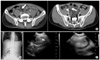
Abdominal radiography showed localized ileus in the lower abdomen, and no free air or any air-fluid level was visible (Fig. 1A). The contrast-enhanced computed tomography (CT) scan showed the target lesion and a complex mass in the ileum with areas of high and low attenuation, which were all suggestive of intussusception. The CT scan also showed a homogeneous mass with fat attenuation in the ileum and this was diagnostic for lipoma (Fig. 1B). Ultrasonography showed the target sign and an echogenic intraluminal mass on the axial scan, and a sausage-shaped lesion was seen on the longitudinal scan (Fig. 1C).
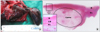
We performed emergency laparotomy under impression of ileo-ileal intussusception that was caused by a lipoma. On laparotomy, an ileo-ileal intussusception was identified 20 cm proximal to the ileocecal valve. Manual reduction was impossible due to the edema of the lesion; the involved ileal segment was resected and an end-to-end anastomosis was performed. Another intussusception was identified at the jejunum 50 cm distal to the ligament of Treitz, and then manual reduction was performed without any difficulty. No mass was palpated in the reduced bowel and no gross abnormality was seen. The postoperative period was uneventful and the patient was discharged on the 13th postoperative day.
DISCUSSION
Intussusception is the invagination of a proximal segment of the bowel with its mesenteric fold (the intussusceptum) into the lumen of the adjacent distal segment (the intussuscipiens) as a result of peristalsis. Since its first description in 1674 by Barbette,(8) it was considered to primarily be a disease of infancy and early childhood. However, many cases that occurred in adults have been reported, and these account for approximately 5% of all intussusceptions, 1% of all intestinal obstructions and it shows an incidence of 0.003 to 0.02% of all hospital admissions.(1-5) Adult intussusception is unusual and it differs from childhood intussusception in its presentation, cause and treatment.
In contrast to childhood intussusception, most of adult intussusceptions are associated with an underlying lesion, and neoplasms, both benign and malignant, are the most frequent causes of adult intussusceptions.(1,2,5-8) In the small bowel, the neoplasms as the leading point of adult intussusceptions are the more often benign, and these include lipoma, Meckel's diverticulum, postoperative adhesion, adenoma and inflammatory fibrous polyps. Approximately 30% of them are malignant lesions, with metastases and lymphoma being the most frequent. In the large bowel, 60% to 70% of the cases show malignant lesions, including adenocarcinoma and lymphoma.(1-3,5,6)
Gastrointestinal lipomas are rare benign tumors that can occur anywhere along the gut, and they are the second most common benign tumors in the small bowel after gastrointestinal stromal tumors.(11) The ileum is the most common site for lipoma in the small bowel, and there has been a study that reviewed such cases and it reported that 83% of the cases were in the ileum and 75% of them were found within 60 cm of ileocecal valve.(11) The peak occurrence is in the fifth to seventh decades of life, with a slight female preponderance. Lipomas are usually solitary and of various sizes ranging from 1 to 30 cm, but multiple lipomas can be found anywhere in the gastrointestinal tract. Because of its usual position immediately superficial to the muscularis propria, gastrointestinal lipoma can produce intussusception as the leading point or intestinal obstruction by occlusion of the bowel lumen.(8-10) Ulceration of the overlying mucosa or intussusception itself can produce gastrointestinal bleeding. The size and location of the lipoma and the mobility afforded by the pseudopedicle, when present, are associated with the clinical signs and symptoms. Lipomas less than 1 cm are usually asymptomatic and they are found incidentally, while 75% of those greater than 4 cm produce symptoms such as intussusception, intestinal obstruction and gastrointestinal bleeding.(8-10) Malignant degeneration has never been reported.
The clinical diagnosis of childhood intussusception is usually suspected before performing imaging studies, yet the diagnosis of adult intussusception is often difficult because of the vague signs and symptoms. Adult intussusception usually presents with nonspecific symptoms that can be acute, intermittent or chronic. Abdominal pain is the most frequent symptom, with or without the symptoms of an intestinal obstruction.(1-3,5,6) Even with the recent advances of the radiologic imaging modalities, intussusception is rarely diagnosed preoperatively. Barussaud et al.(2) reported that the preoperative diagnosis was made in only 52% of patients, Azar and Berger(1) reported 32%, and Nagorney et al.(6) reported 35%. Computed tomography (CT) is the imaging method of choice for diagnosing intussusception and it can helpful in revealing the underlying lesion,(1-5) and some series have reported that CT scanning allows making a correct preoperative diagnosis in up to 80% of the cases, although barium enema can also help make the diagnosis for colonic intussusceptions.(1) The CT findings of intussusception are a mass-like lesion, including the outer intussuscipiens, the inner intussusceptum and an eccentric fat density mass that represents the intussuscepted mesenteric fat, and this appears as a "target" or a "sausage" mass according to the cut axis.(3,4) Our patient had the typical findings of intussusception on CT and the CT scan showed a homogeneous intraluminal mass with fat attenuation (Hounsfield units between -80 and -120) in the ileum, so we could suspect the diagnosis of a ileo-ileal intussusception that was caused by an ileal lipoma.(9-11)
Once the diagnosis of intussusception in adults is made, surgical intervention is indicated and surgical resection remains the recommended treatment for nearly all cases because most of adult intussusceptions have an underlying structural lesion, and adult intussusceptions have a relatively high incidence of malignancy. However, there is a controversy about whether or not the intussusception should be initially reduced before resection. Nagorney et al.(6) reported that two-thirds of the colonic intussusceptions were associated with primary carcinoma of the colon and one-third of the enteric intussusceptions were related to underlying malignancies, but the incidence of primary malignant lesions of the small bowel was low compared with the more frequent metastatic lesions. So, they proposed that resection without initial surgical reduction is favored for the colonic intussusceptions, and initial reduction followed by limited surgical resection is the preferred treatment for the enteric intussusceptions. Azar and Berger(1) reported that about half of the enteric intussusceptions were related to underlying malignancies and about half of the other benign enteric intussusceptions were associated with postoperative factors. They proposed that all the patients with colonic intussusception and all the patients with enteric intussusception who have not had a previous laparotomy should undergo resection without initial reduction. Yet the most widely accepted view is that resection without reduction should be performed for most cases of colonic intussusception because of the high incidence of underlying malignancy. Further, initial surgical reduction should be attempted for the cases of enteric intussusception, provided there is no sign of inflammation or bowel ischemia and a malignant lesion is not suspected, to avoid unnecessary excision of healthy bowel.(2,3,7,8,11) In recent years, with increasing exact preoperative diagnosis of an intussusception and an underlying lesion according to the CT scan, several studies have proposed the laparoscopic approach as a safe, feasible therapeutic option for selected cases of adult intussusceptions, although the role of laparoscopy in managing adult intussusceptions is not yet clearly defined.(12-14)
Preoperatively, we presumed that our patient was suffering from an ileo-ileal intussusception caused by a lipoma of the ileum according to the typical CT findings, and we tried to initially reduce the intussusception during laparotomy. But manual reduction was impossible due to the edema of the lesion, and an ileum of some length had to be resected.
Adult intussusception is rare and even with the recent advances of the radiologic imaging modalities, intussusception is rarely diagnosed preoperatively. Yet for some cases, making the preoperative diagnosis of intussusception and revealing an underlying lesion are possible from the CT scan or from other imaging modalities, and so these imaging modalities can help to plan a surgical approach such as initial reduction or resection.
 XML Download