

 PDF
PDF ePub
ePub Citation
 Print
Print


Abstract
Purpose
Left iliac vein compression is a risk factor for deep vein thrombosis (DVT) and often can be symptomatic. We wanted to know the incidence of left iliac vein compressions in the general population and the relationship between iliac vein compression and outflow fraction of the lower extremities.
Methods
1,523 cases examined with abdomen - pelvis CT were included in this study. Left iliac vein compression was calculated as the diameter of left common iliac vein at the site of maximal compression divided by the diameter of the uncompressed caudal common iliac vein. These cases were divided into 4 groups by the degree of iliac vein compression. In addition, left lower extremity venous outflow fractions in 106 patients with abdomen - pelvis CT images were recorded and analyzed.
Results
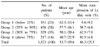
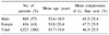
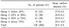
The number and mean age of each group (group 1, 2, 3 and 4) were 351 (23%) & 62.3±13.4, 426 (28%) & 57.7±16.1, 529 (35%) & 50.1±20.4 and 217 (14%) & 40.7±22.9, respectively. While the age of patients was decreasing, the occurrence of left iliac vein compression increased (P<0.01). The mean left low extremity venous outflow fractions of each group (group 1, 2, 3 and 4) was 27.9±6.9%, 26.9±6.4%, 25.9±6.7% and 19.1±6.6%, respectively. The mean outflow fraction of group 4 was significantly lower than that of other groups (P<0.01). There was a tendency that the more left iliac vein compression increased, the more outflow fraction decreased (P=0.011).
Figures and Tables
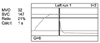 | Fig. 1Example of outflow fraction. MVO = maximum venous outflow per 1 second, equal to V1; SVC = systemic venous capacitance, equal to V; Ratio = equal to outflow fraction.
|
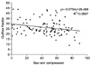
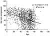 | Fig. 3Correlation of age and Lt. iliac vein compression. Pearson's correlation is -0.363 between age and iliac vein compression (P<0.01) (R2=-0.1318).
|
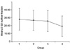 | Fig. 4The mean outflow fraction of group 4 was significantly lower than that of other groups (P<0.01).
|
References
1. May R, Thurner J. The cause of the predominantly sinistral occurrence of thrombosis of the pelvic veins. Angiology. 1957. 8:419–427.
2. Cockett FB, Thomas ML. The iliac compression syndrome. Br J Surg. 1965. 52:816–821.
3. Rutherford RB. Vascular Surgery. 2005. 6th ed. Philadelphia: Elsevier Saunders;230.
4. Kibbe MR, Ujiki M, Goodwin AL, Eskandari M, Yao J, Matsumura J. Iliac vein compression in an asymptomatic patient population. J Vasc Surg. 2004. 39:937–943.
5. Oguzkurt L, Tercan F, Pourbagher MA, Kizilkilic O, Turkoz R, Boyvat F. Computed tomography findings in 10 cases of iliac vein compression (May-Thurner) syndrome. Eur J Radiol. 2005. 55:421–425.
6. Moreland NC, Ujiki M, Matsumura JS, Morasch MD, Eskandari MK, Pearce WH, et al. Decreased incidence of left common iliac vein compression in patients with abdominal aortic aneurysms. J Vasc Surg. 2006. 44:595–600.
7. Hurst DR, Forauer AR, Bloom JR, Greenfield LJ, Wakefield TW, Williams DM. Diagnosis and endovascular treatment of iliocaval compression syndrome. J Vasc Surg. 2001. 34:106–113.
8. Criado E, Farber MA, Marston WA, Daniel PF, Burnham CB, Keagy BA. The role of air plethysmography in the diagnosis of chronic venous insufficiency. J Vasc Surg. 1998. 27:660–670.
9. Owens LV, Farber MA, Young ML, Carlin RE, Criado-Pallares E, Passman MA, et al. The value of air plethysmography in predicting clinical outcome after surgical treatment of chronic venous insufficiency. J Vasc Surg. 2000. 32:961–968.
10. Oguzkurt L, Ozkan U, Ulusan S, Koc Z, Tercan F. Compression of the left common iliac vein in asymptomatic subjects and patients with left iliofemoral deep vein thrombosis. J Vasc Interv Radiol. 2008. 19:366–371.
11. Fraser DG, Moody AR, Martel A, Morgan PS. Re-evaluation of iliac compression syndrome using magnetic resonance imaging in patients with acute deep venous thromboses. J Vasc Surg. 2004. 40:604–611.
12. Chung JW, Yoon CJ, Jung SI, Kim HC, Lee W, Kim YI, et al. Acute iliofemoral deep vein thrombosis: evaluation of underlying anatomic abnormalities by spiral CT venography. J Vasc Interv Radiol. 2004. 15:249–256.
 XML Download