

 PDF
PDF ePub
ePub Citation
 Print
Print


Abstract
Primary angiosarcoma of the spleen is an extremely rare malignancy, the pathogenesis of which is not completely understood, with high metastatic potential and an exceedingly poor prognosis, regardless of treatment regimen. The major complication is splenic rupture, which often leads to fatal hemoperitoneum. Overall, since 1879 when Langerhans described the first case of angiosarcoma of the spleen, there have been approximately 200 cases reported in the literature. Moreover, to the best of our knowledge, spontaneous rupture of primary splenic angiosarcoma and spontaneous rupture of remnant or recurred angiosarcoma is extremely rare, and no cases were reported in English literature. We report a case of spontaneous splenic rupture due to angiosarcoma in a 68-year-old man, and also review the existing literature.
Figures and Tables
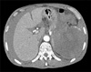 | Fig. 1The abdominal CT scan. Abdominal CT scan revealed a large amount of hemoperitoneum due to splenic rupture, and hematoma, which replaces splenic parenchyma.
|
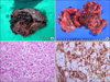 | Fig. 2Gross and microscopic finding. (A, B) Hemorrhagic and nodular lesions are seen in ruptured spleen. (C) Microscopic findings show presence of sinusoidal vascular spaces lined by large pleomorphic tumor cells with irregular hyperchromatic nuclei and abundant eosinophilic cytoplasm. (H&E stain, ×200). (D) Immunoreactivity of splenic angiosarcoma for CD31 is seen (×200).
|
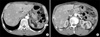 | Fig. 3The follow-up abdominal CT scans (36th postoperative day). There is seen an angiomatous mass lesion (angiosarcoma, maximum 3×4 cm sized, white arrow) in splenectomy bed (A), with multiple hypervascular metastases in the liver (A, B).
|
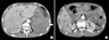 | Fig. 4The follow-up abdominal CT scans (78th postoperative day). About two months later, the follow-up abdominal CT scan revealed a large amount of hematoma, possibly due to rupture of recurred mass lesion (angiosarcoma, maximum 10×9 cm sized, white arrow) in splenectomy bed (A), and more aggravation of multiple hypervascular metastases in the liver (A, B).
|
References
1. Manouras A, Giannopoulos P, Toufektzian L, Markogiannakis H, Lagoudianakis EE, Papadima A, et al. Splenic rupture as the presenting manifestation of primary splenic angiosarcoma in a teenage woman: a case report. J Med Case Reports. 2008. 2:133.
2. Buckner JW 3rd, Porterfield G, Williams GR. Spontaneous splenic rupture secondary to angiosarcoma. J Okla State Med Assoc. 1990. 83:211–213.
3. Montemayor P, Caggiano V. Primary hemangiosarcoma of the spleen associated with leukocytosis and abnormal spleen scan. Int Surg. 1980. 65:369–373.
4. Falk S, Krishnan J, Meis JM. Primary angiosarcoma of the spleen. A clinicopathologic study of 40 cases. Am J Surg Pathol. 1993. 17:959–970.
5. Neuhauser TS, Derringer GA, Thompson LD, Fanburg-Smith JC, Miettinen M, Saaristo A, et al. Splenic angiosarcoma: a clinicopathologic and immunophenotypic study of 28 cases. Mod Pathol. 2000. 13:978–987.
6. Keymeulen K, Dillemans B. Epitheloid angiosarcoma of the splenic capsula as a result of foreign body tumorigenesis. A case report. Acta Chir Belg. 2004. 104:217–220.
7. Al'pidovskii VK, Suvorova EV, Halil MA. Angiosarcoma of the spleen with consumption coagulopathy. Ter Arkh. 1990. 62:124–126.
8. Kinoshita T, Ishii K, Yajima Y, Sakai N, Naganuma H. Splenic hemangiosarcoma with massive calcification. Abdom Imaging. 1999. 24:185–187.
9. Safapor F, Aghajanzade M, Kohsari M, Hoda S, Safarpor D. Spontaneous rupture of the spleen: a case report and review of the literature. Saudi J Gastroenterol. 2007. 13:136–137.
10. Naka N, Ohsawa M, Tomita Y, Kanno H, Uchida A, Myoui A, et al. Prognostic factors in angiosarcoma: a multivariate analysis of 55 cases. J Surg Oncol. 1996. 61:170–176.
 XML Download