

 PDF
PDF ePub
ePub Citation
 Print
Print


Abstract
Purpose
FK506 (tacrolimus) is a widely used immunosuppressive agent in the treatment of various medical conditions, including autoimmune disease, bone marrow and organ transplantations. Previously FK506 was known to cause apoptotic death of human Jurkat T cells.
Methods
The current study was designed to analyze the gene expression pattern of Jurkat T cells after FK506 application by using cDNA microarray. Treatment of Jurkat T cells with FK506 resulted in a decrease of cell viability in a time- and dose-dependent manner. Next, total RNA of Jurkat T cells was extracted by using TRIzol reagent and used to carry out a confirmation test for the purity and integrity of total RNA.
Results
Gene expression levels related to apoptosis and cell cycle process were mainly focused to analyze in FK506-treated Jurkat T cells. According to the inhibition of calcineurin activity, MARCKS in PKC substrates and Sp3 transcription factor was markedly increased in FK506-treated cells. Also, cell cycle control gene Id1 and Id3 were induced in expression from FK506-treated Jurkat T cells. However, FK506 decreased the expression of Src homology 2, G protein, and MEK 2 genes in bioactive peptide induced signaling pathway. It also reduced the expression level of the insulin receptor, DRPLA and Bai1-associated protein 2 genes, which are involved in the regulation of cell motility and morphology control.
Figures and Tables
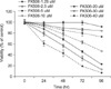 | Fig. 1FK506 decreased the viability of Jurkat cells in dose- and time-dependent manners. Cells were treated with various concentration of FK506 for 96 hours, then cell viability was measured by MTT assay after FK506 treatment. Data represent the mean±standard deviation (SD) of triplicates.
|
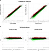 | Fig. 2Modulation of gene expression following FK506-treated Jurkat cells. Total RNA was prepared from Jurkat using TRIzol as described by manufacturer. (A) Scatter plot analysis. The x and y axes represent Cy3 (Control) and Cy5 (FK506 10µM or 20µM) signal intensity values, respectively. These represent up-regulated (upper lines; red spots) or down-regulated (under lines; green spots) genes. (B) MA plot analysis. The x and y axes represent A (R+G/2) and M (R/G) signal intensity values. The R and G repesent Cy5-background and Cy3-background signal intensity, respectively. These represent up-regulated (upper lines; red spots) or down-regulated (under lines; green spots) genes.
|


References
1. Pridohl O, Heinemann K, Hartwig T, Witzigmann H, Lamesch P, Fangmann J, et al. Low-dose immunosuppression with FK 506 and sirolimus after liver transplantation: 1-year results. Transplant Proc. 2001. 33:3229–3231.
2. Harding MW, Galat A, Uehling DE, Schreiber SL. A receptor for the immunosuppressant FK506 is a cis-trans peptidyl-prolyl isomerase. Nature. 1989. 341:758–760.
3. Maki N, Sekiguchi F, Nishimaki J, Miwa K, Hayano T, Takahashi N, et al. Complementary DNA encoding the human T-cell FK506-binding protein, a peptidylprolyl cis-trans isomerase distinct from cyclophilin. Proc Natl Acad Sci U S A. 1990. 87:5440–5443.
4. Brown EJ, Albers MW, Shin TB, Ichikawa K, Keith CT, Lane WS, et al. A mammalian protein targeted by G1-arresting rapamycin-receptor complex. Nature. 1994. 369:756–758.
5. Bagnis C, Deray G, Dubois M, Adabra Y, Jacquiaud C, Jaudon MC, et al. Comparative acute nephrotoxicity of FK-506 and ciclosporin in an isolated in situ autoperfused rat kidney model. Am J Nephrol. 1997. 17:17–24.
6. Olyaei AJ, de Mattos AM, Bennett WM. Nephrotoxicity of immunosuppressive drugs: new insight and preventive strategies. Curr Opin Crit Care. 2001. 7:384–389.
7. Weiss A, Wiskocil RL, Stobo JD. The role of T3 surface molecules in the activation of human T cells: a two-stimulus requirement for IL 2 production reflects events occurring at a pre-translational level. J Immunol. 1984. 133:123–128.
8. Reem GH, Cook LA, Vilcek J. Gamma interferon synthesis by human thymocytes and T lymphocytes inhibited by cyclosporin A. Science. 1983. 221:63–65.
9. Arbuzova A, Schmitz AA, Vergeres G. Cross-talk unfolded: MARCKS proteins. Biochem J. 2002. 362:1–12.
10. Hagen G, Muller S, Beato M, Suske G. Sp1-mediated transcriptional activation is repressed by Sp3. EMBO J. 1994. 13:3843–3851.
11. Ge Y, Jensen TL, Matherly LH, Taub JW. Physical and functional interactions between USF and Sp1 proteins regulate human deoxycytidine kinase promoter activity. J Biol Chem. 2003. 278:49901–49910.
12. Ko JL, Liu HC, Loh HH. Role of an AP-2-like element in transcriptional regulation of mouse mu-opioid receptor gene. Brain Res Mol Brain Res. 2003. 112:153–162.
13. Rotheneder H, Geymayer S, Haidweger E. Transcription factors of the Sp1 family: interaction with E2F and regulation of the murine thymidine kinase promoter. J Mol Biol. 1999. 293:1005–1015.
14. Riechmann V, van Cruchten I, Sablitzky F. The expression pattern of Id4, a novel dominant negative helix-loop-helix protein, is distinct from Id1, Id2 and Id3. Nucleic Acids Res. 1994. 22:749–755.
15. Zebedee Z, Hara E. Id proteins in cell cycle control and cellular senescence. Oncogene. 2001. 20:8317–8325.
16. Yokota Y. Id and development. Oncogene. 2001. 20:8290–8298.
17. Tewari M, Beidler DR, Dixit VM. CrmA-inhibitable cleavage of the 70-kDa protein component of the U1 small nuclear ribonucleoprotein during Fas- and tumor necrosis factor-induced apoptosis. J Biol Chem. 1995. 270:18738–18741.
18. Casciola-Rosen LA, Anhalt GJ, Rosen A. DNA-dependent protein kinase is one of a subset of autoantigens specifically cleaved early during apoptosis. J Exp Med. 1995. 182:1625–1634.
19. Lazebnik YA, Takahashi A, Moir RD, Goldman RD, Poirier GG, Kaufmann SH, et al. Studies of the lamin proteinase reveal multiple parallel biochemical pathways during apoptotic execution. Proc Natl Acad Sci U S A. 1995. 92:9042–9046.
20. Mashima T, Naito M, Fujita N, Noguchi K, Tsuruo T. Identification of actin as a substrate of ICE and an ICE-like protease and involvement of an ICE-like protease but not ICE in VP-16-induced U937 apoptosis. Biochem Biophys Res Commun. 1995. 217:1185–1192.
21. Brancolini C, Benedetti M, Schneider C. Microfilament reorganization during apoptosis: the role of Gas2, a possible substrate for ICE-like proteases. EMBO J. 1995. 14:5179–5190.
22. Nishimori H, Shiratsuchi T, Urano T, Kimura Y, Kiyono K, Tatsumi K, et al. A novel brain-specific p53-target gene, BAI1, containing thrombospondin type 1 repeats inhibits experimental angiogenesis. Oncogene. 1997. 15:2145–2150.
23. Kaur B, Brat DJ, Devi NS, Van Meir EG. Vasculostatin, a proteolytic fragment of brain angiogenesis inhibitor 1, is an antiangiogenic and antitumorigenic factor. Oncogene. 2005. 24:3632–3642.
24. Bjarnadottir TK, Fredriksson R, Hoglund PJ, Gloriam DE, Lagerstrom MC, Schioth HB. The human and mouse repertoire of the adhesion family of G-protein-coupled receptors. Genomics. 2004. 84:23–33.
25. Park D, Tosello-Trampont AC, Elliott MR, Lu M, Haney LB, Ma Z, et al. BAI1 is an engulfment receptor for apoptotic cells upstream of the ELMO/Dock180/Rac module. Nature. 2007. 450:430–434.
26. Cho HJ, Shashkin P, Gleissner CA, Dunson D, Jain N, Lee JK, et al. Induction of dendritic cell-like phenotype in macrophages during foam cell formation. Physiol Genomics. 2007. 29:149–160.
 XML Download