

 PDF
PDF ePub
ePub Citation
 Print
Print


INTRODUCTION
Gallbladder torsion is defined as the rotation of the gallbladder on its mesentery along the axis of the cystic duct and cystic artery; this disease is known to be related with a mobile gallbladder and variations of the mesenteric anatomy. It was first described in 1898 by Wendel(1) and approximately 500 or more cases have currently been reported in the literature.(2-4) Even with the recent advances in the radiologic imaging modalities, gallbladder torsion is a rare disease. The symptoms of this disease are largely non-specific, so making the preoperative diagnosis of gallbladder torsion is difficult, and the definite diagnosis is usually made at the time of surgery. Early diagnosis and prompt cholecystectomy for treating this disease are important in order to avoid the complications of gangrene and perforation, and to reduce the resultant mortality.(5)
We present here a case of gallbladder torsion with accompanying acute appendicitis and we review the clinical aspects of this disease. We missed the diagnosis of gallbladder torsion preoperatively and the definite diagnosis was made at the time of surgery, so we now review and correlate the CT findings with the surgical findings.
CASE REPORT
An 89-year-old woman presented to the emergency room of our hospital with a 1-day history of severe epigastric pain. She had a past history of bronchiectasis and compression fracture of the 1st lumbar vertebra. The epigastric pain had started intermittently along with nausea and vomiting and she had failed to pass faeces for 4 days prior to presentation. The physical examination revealed a thin elderly woman (height: 150 cm, body weight: 40 kg) with a blood pressure of 130/90 mmHg, a pulse of 92 beats/min and a body temperature of 36.5℃. The abdomen was not distended, but there was tenderness in the right side, and especially in the right upper quadrant. The bowel sounds were not increased. The laboratory findings showed a white blood cell count of 14,400/mm3, a hemoglobin level of 12.6 g/dl, a hematocrit of 37.6% and the platelet count was 316,000/mm3. All the other studies, including the electrolytes and urinalysis, were within the reference limits. Abdominal radiography showed localized ileus, and no free air or an air fluid level was visible. She was admitted and treated with IV fluid and analgesics.
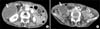
On post-admission day 1, her abdominal pain increased in intensity and it then became constant. The physical examination revealed a body temperature of 38.0℃ and there was tenderness and marked guarding in the right side of the abdomen, and laboratory findings showed a white blood cell count of 22,900/mm3. A contrast-enhanced computed tomography (CT) scan was performed; this showed a markedly distended gallbladder with a thickened wall that suggested acute cholecystitis and there was an inflamed appendix in the retrocecal area. A dilated extrahepatic bile duct was noted, but no definite obstructing lesion was found (Fig. 1). Ultrasonography was performed and this showed a markedly distended gallbladder with a thickened wall with a sonographic Murphy's sign that suggested acute cholecystitis, but no stones were seen.
We performed emergency laparotomy through a midline incision under the impression of acute acalculous cholecystitis and acute appendicitis. At laparotomy, a very large, distended and gangrenous gallbladder was found in the upper abdomen, and a distended, edematous appendix was found in the lower abdomen. The gallbladder was not attached to the liver bed and it displayed 360 degree torsion around the junction of the neck of the gallbladder and cystic duct (Fig. 2). Detorsion was done and cholecystectomy was easily performed. Appendectomy was performed as well. Operative cholangiography was done through the cystic duct and this showed a dilated extrahepatic bile duct, but no filling defect. The postoperative period was uneventful and the patient was discharged on the 11th postoperative day.
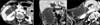
Unfortunately, the preoperative diagnosis of gallbladder torsion was missed, so we reviewed and correlated CT findings with the surgical findings. The contrast-enhanced CT scan showed a tapered cystic duct that had no obvious continuation with the gallbladder lumen, and crowded mesenteric and cystic arteries were seen with a retracted contour of the medial border of the S5 segment of the liver (Fig. 3A). The coronal reformatted image of the CT scan showed a floating, markedly dilated gallbladder lumen with a diffusely thickened wall and a poorly enhanced mucosal layer of the gallbladder neck (Fig. 3B). The curved multiplanar reformation image showed twisted mesenteric and cystic arteries with a retracted liver surface of the S5 segment of the liver and an inflamed appendix in the retrocecal area (Fig. 3C).
The resected gallbladder was about 12.5 cm in length and it had a thickened edematous wall with gangrenous change. There were no stones in the gallbladder lumen. The microscopic findings of the gallbladder were consistent with acute gangrenous cholecystitis (Fig. 4A). The resected appendix was about 6 cm in length and 1.5 cm in diameter and the wall was edematous. The microscopic findings of the appendix were consistent with acute appendicitis (Fig. 4B).
DISCUSSION
Gallbladder torsion is a rare disease. Since its first description in 1898 by Wendel,(1) approximately 500 or more cases have been reported in the literature.(2-4) Although this disease can occur at any age, 85% of the reported cases have occurred between the ages of 60 and 80 years and there is a female preponderance with a female to male ratio of 3:1.(3,4) The incidence of this malady appears to have recently increased, and this is possibly related to the increased life expectancy.(6,7)
The etiology of gallbladder torsion is unknown. However, several factors have been postulated to play causative roles. There are two anatomical variations in the attachment of the gallbladder to the liver to allow mobility of the gallbladder along the axis of the cystic duct and the cystic artery. These anatomical variations include a long mesentery and a very short or absent mesentery, which allow the gallbladder to float and this results in torsion. (3-9) The long mesentery type of anatomical variation of the gallbladder can be acquired with aging. Atrophy of the liver and the loss of both visceral fat and elasticity with advancing age contribute the mobility of the gallbladder. (3-9) Other precipitating factors are also necessary to initiate torsion, include kyphoscoliosis of the spine, arteriosclerosis of the cystic artery and intense peristalsis of neighboring organs.(3-9) Gallstones are present in 20~50% of the cases, but they are unlikely to be the cause of gallbladder torsion.(4,6-8) Our patient in this case was a thin elderly woman (height: 150 cm, body weight: 40 kg, BMI (body mass index): 17.8) and she had a short mesentery (a floating gallbladder), but the role of the acute appendicitis for this patient's gallbladder torsion is unknown.
The symptoms of gallbladder torsion are largely nonspecific and they mimic those of acute cholecystitis.(3-9) When the rotation is less than 180 degrees (incomplete), the pain is similar to that of biliary colic, but when the rotation is more than 180 degrees (complete) and the blood supply to the gallbladder is compromised, then the pain is similar to that of acute cholecystitis. The physical examination usually shows non-specific findings, such as tenderness, guarding and low-grade fever, which mimic those findings of acute cholecystitis.(3-9) The gallbladder may or may not be palpated. These non-specific symptoms and the non-specific findings on physical examination and its rarity make the preoperative diagnosis difficult. Lau et al.(10) proposed 3 triads for the clinical diagnosis of gallbladder torsion, and these include specific symptoms (a short history, abdominal pain and early vomiting), physical signs (abdominal mass, the absence of toxaemia and a pulse rate-temperature discrepancy) and physical characteristics (thin, elderly and a deformed spine). The laboratory evaluations are not helpful in most cases, and they usually show a normal or elevated white cell count and normal liver function tests.
Even with the recent advances of the radiologic imaging modalities, making the correct preoperative diagnosis of gallbladder torsion is difficult and most cases are diagnosed at the time of surgery. However, ultrasonography and computed tomography (CT) are useful modalities and some findings of ultrasonography and CT can help clinicians diagnose gallbladder torsion preoperatively when they have a suspicion of this disease.(2,11-13) The specific findings of gallbladder torsion include a large anteriorly floating gallbladder without gallstones and an echogenic conical structure in the neck with discontinuity of the lumen. The non-specific findings include gross wall thickening and distension, and these findings can also be present in both torsion and calculous cholecystitis. Kim et al.(14) emphasized the whirlpool-like flow signals of vessels in the twisting knot ("whirlpool sign") on Doppler ultrasonography. Magnetic resonance cholangiopancreatography (MRCP) may be useful in diagnosing gallbladder torsion and this can show a V-shaped distortion of the extrahepatic bile ducts due to traction by the cystic duct, tapering and a twisting interruption of the cystic duct, a distended and enlarged gallbladder that is deviated to the midline of the abdomen and a difference in intensity between the gallbladder and the extrahepatic bile ducts and the cystic duct.(15)
The correct preoperative diagnosis of gallbladder torsion was missed in this case, so we reviewed and correlated the CT findings with the surgical findings. Retrospectively, the specific CT findings that suggested gallbladder torsion in our case include a tapered cystic duct that had no obvious continuation with the gallbladder lumen, crowded mesenteric and cystic arteries with a retracted contour of the medial border of the S5 segment of the liver, a floating, markedly dilated gallbladder lumen with a diffusely thickened wall and a poorly enhanced mucosal layer of the gallbladder neck on the coronal reformatted image, and twisted mesenteric and cystic arteries with a retracted liver surface of the S5 segment on the curved multi-planar reformation image. If we had had a suspicion of gallbladder torsion on the basis of the clinical situation (the patient was a thin elderly woman with symptoms that were consistent with acute cholecystitis and she responded poorly to conservative management, and there were no gallstones seen on ultrasonography and CT), the correct preoperative diagnosis might have been possible, based on the specific CT findings.
Emergency cholecystectomy is required for treating gallbladder torsion, and the laparoscopic approach is now recommended as the first choice of treatment due to the increased experience with laparoscopic cholecystectomy. (3,4,6,9) An early diagnosis and performing prompt cholecystectomy for this disease is important in order to avoid the complications of gangrene and perforation, and to reduce the resultant mortality.
In conclusion, gallbladder torsion is a rare clinical entity that requires immediate surgical treatment, but making the correct preoperative diagnosis is difficult and most cases are diagnosed at the time of surgery. We experienced a case of an 89-year-old woman with gallbladder torsion and accompanying acute appendicitis. A high index of suspicion on the basis of the clinical situation is important and the specific CT findings are useful for making the correct preoperative diagnosis of gallbladder torsion. In our case, the role of the acute appendicitis for this patient's gallbladder torsion is unknown.


 XML Download