Abstract
Duplicated inferior vena cava (IVC) is a congenital anomaly seen rarely in the general population. Patients with IVC variants usually do not present any symptoms and are found incidentally in many cases. However, physicians are urged to recognize the presence of such anomalies during diagnostic or invasive procedures as these variants of blood vessel systems can impose substantial implications in certain clinical situations. Subsequently, information about IVC variants may become critical if surgical injuries or predisposing conditions act as life-threatening risks to patients during medical procedures. We present a case of duplicated IVC in a 68?year?old female patient with rectal cancer where an IVC anomaly was found during surgical resection of her tumor. From our experience, we emphasize the importance of having the knowledge of IVC variations in patients undergoing invasive surgical procedures which may involve large vessels.
Figures and Tables
 | Fig. 1Operative finding of duplicated inferior vena cava (IVC). Right and left IVC run along both sides of the aorta. R = right IVC; L = left IVC; A = aorta. |
 | Fig. 2CT images of 68-year-old female with duplicated inferior vena cava. (A) Contrast enhanced CT shows a coronal image of duplicated inferior vena cava. Left inferior vena cava drains into the left renal vein (thick white arrow) and then joined the right inferior vena cava. (B) Contrast enhanced CT shows an axial image of duplicated inferior vena cava (thin white arrows). |
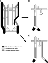
 | Fig. 3Diagrams show the development of inferior vena cava. (A) Inferior vena cava develops from three pairs of veins, posterior cardinal vein, subcardinal vein and supracardinal vein. (B) Development of normal inferior vena cava. (C) Development of duplicated inferior vena cava. *LGV = left gonadal vein. |
References
1. Friedland GW, deVries PA, Nino-Murcia M, King BF, Leder RA, Stevens S. Congenital anomalies of the inferior vena cava: embryogenesis and MR features. Urol Radiol. 1992. 13:237–248.
2. Garcia-Fuster MJ, Forner MJ, Flor-Lorente B, Soler J, Campos S. Inferior vena cava malformations and deep venous thrombosis. Rev Esp Cardiol. 2006. 59:171–175.
3. Koc Z, Ulusan S, Oguzkurt L, Tokmak N. Venous variants and anomalies on routine abdominal multi-detector row CT. Eur J Radiol. 2007. 61:267–278.
4. Evans JC, Earis J, Curtis J. Thrombosed double inferior vena cava mimicking paraaortic lymphadenopathy. Br J Radiol. 2001. 74:192–194.
5. Arisawa C, Kihara K, Fujii Y, Ishizaka K, Masuda H, Oshima H. Possible misinterpretation on computed tomography of left inferior vena cava as retroperitoneal lymph node metastasis: a report of two cases. Int J Urol. 1999. 6:215–218.
6. Shingleton WB, Hutton M, Resnick MI. Duplication of inferior vena cava: its importance in retroperitoneal surgery. Urology. 1994. 43:113–115.
7. Lee CH, Jang LC, Park JW, Choi JW. The incidence of inferior vena cava anomalies by MDCT. J Korean Surg Soc. 2008. 74:60–64.
8. Reinus WR, Gutierrez FR. Duplication of the inferior vena cava in thromboembolic disease. Chest. 1986. 90:916–918.
9. Kouroukis C, Leclerc JR. Pulmonary embolism with duplicated inferior vena cava. Chest. 1996. 109:1111–1113.
10. Sartori MT, Zampieri P, Andres AL, Prandoni P, Motta R, Miotto D. Double vena cava filter insertion in congenital duplicated inferior vena cava: a case report and literature review. Haematologica. 2006. 91:ECR30.


 PDF
PDF ePub
ePub Citation
 Print
Print


 XML Download