

 PDF
PDF ePub
ePub Citation
 Print
Print


INTRODUCTION
Duodenal anomalies, such as duodenal atresia, intraluminal webs, annular pancreas, have been recognized as primary pediatric surgical disease. Occasionally there are instances that remain asymptomatic until adulthood.
Intraluminal duodenal diverticulum (IDD), so called "windsock web", is a rare, recognized congenital condition in the adult. Besides the common symptoms of epigastric discomfortness, nausea and vomiting, sometimes it presents with an obstruction, pancreatitis, and bleeding.(1) The diagnosis is usually made by an upper gastrointestinal contrast study and a gastroduodenoscopy. We report a case found by gastroduodenoscopy evaluated for intermittent melena and successfully managed surgically.
CASE REPORT
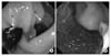
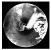
A 32-year-old man complaining an upper abdominal discomfortness and intermittent melena visited the Department of Medicine in our institution, he had no history of health problems and medications. Laboratory tests didn't show any other abnormal findings but a mild anemia with an Hb level of 10.2 g/dl. A fiberoptic gastroduodenoscopy and EUS revealed a cystic mass at the 2nd portion of duodenum which needs making a differentiation from a duplication cyst of duodenum and a diverticulum. A close examination found an opening of the mass located below an ampulla of Vater distinguishing from a true lumen of the duodenum (Fig. 1). There was a shallow ulcer around the opening thought to be a focus of an intermittent melena. And subsequent examinations, an upper gastrointestinal series and an abdominal CT, revealed an intraluminal contrast filling with a radiolucent halo and a sac-like projection mimicking an intussusception between 2nd and 3rd portion of the duodenum (Fig. 2, 3). The patient was referred to our department for surgical correction.
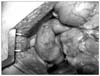
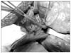
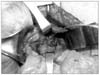
At exploration, there were no abnormal findings, grossly (Fig. 4). Kocher maneuver was performed for mobilizing the duodenum, and then a longitudinal duodenotomy, about 2 cm, was made through the anterior wall between the second and the third portion. We could identify an elongated, sac-like mucosal fold with other opening just distal to the ampulla of Vater along the posterior wall. To avoid an injury to the ampulla of Vater, inserted a catheter to its opening, and then tried to make an eversion of the fold for adequate excision (Fig. 5).
Mucosectomy was performed circumferentially around the fold for excision. Repaired a defected mucosa as a usual maneuver, interrupted absorbable suture (Fig. 6). Finished the procedure after closing the duodenotomy with an interrupted absorbable suture through a transverse axis.
DISCUSSION
An intraluminal duodenal diverticulum is thought to be one of defects in duodenal development, with a variety of duodenal web, stenosis and atresia.(2) It forms as result of a failure in full recanalization and a membraneous tissue stretching across a portion of the duodenal lumen, so the peristaltic movement of the duodenum could make formation of a sac-like structure, intraluminal duodenal diverticulum.(3) This point is a difference between an intraluminal duodenal diverticulum and a duodenal duplication which is composed of a duodenal mucosal lining and a smooth muscular layer. There have been less than 150 cases reported in the literature worldwide up to now.(4)
It usually found at the second portion of the duodenum and arises near the ampulla of Vater, but rarely at the third portion of the duodenum.
Clinically this condition is asymptomatic in the most cases and rarely appear until the third decade of life, however may give rise to some symptoms associated with complications, such as upper gastrointestinal bleeding, obstruction, acute and recurrent pancreatitis.(5) Gastrointestinal bleeding and acute pancreatitis are associated with intraluminal duodenal diverticulum in approximately 20~25% of the reported cases. Especially, possible causes of bleeding are thought to be an ulceration of diverticulum and a trauma induced by food passage. As in this case, we could find ulceration at the base of large diverticulum near the ampulla of Vater which might be a cause of bleeding.(6,7)
Diagnosis is usually made by an upper gastrointestinal series showing a finger-like sac filled with contrast and a radiolucent halo formed by the surround mucosa. In some cases, the CT findings of intraluminal diverticulum, a low-density flap within the contrast-filled duodenum mimicking an intussusception were reported.(8,9) Nowadays, a gastroduodenal endoscopy is a preferred tool for gastrointestinal problems such as a bleeding, so that it could plays an important role in the diagnosis and management for this condition.(10)
Although there were some reports about successful endoscopic excision, the management of patients with a symptomatic intraluminal duodenal diverticulum has been primarily surgical, the procedure of choice is a duodenotomy and excision of the diverticulum.(1) When performing this procedure, a careful search for the biliary and pancreatic orifice is essential before excision of the diverticulum to avoid injury to the ampulla of Vater.
In summary, intraluminal duodenal diverticulum is an infrequent cause of gastrointestinal bleeding, however may be considered in the case of subtle gastrointestinal hemorrhage with or without anemia at the adulthood who had no medical problems before. The preferred management of intraluminal duodenal diveticulum is surgical excision with safety and effectiveness.

 XML Download