Abstract
Purpose
To report a case of retinopathy in a patient with chronically resistant idiopathic thrombocytopenic purpura (ITP) associated with a poor prognosis.
Case summary
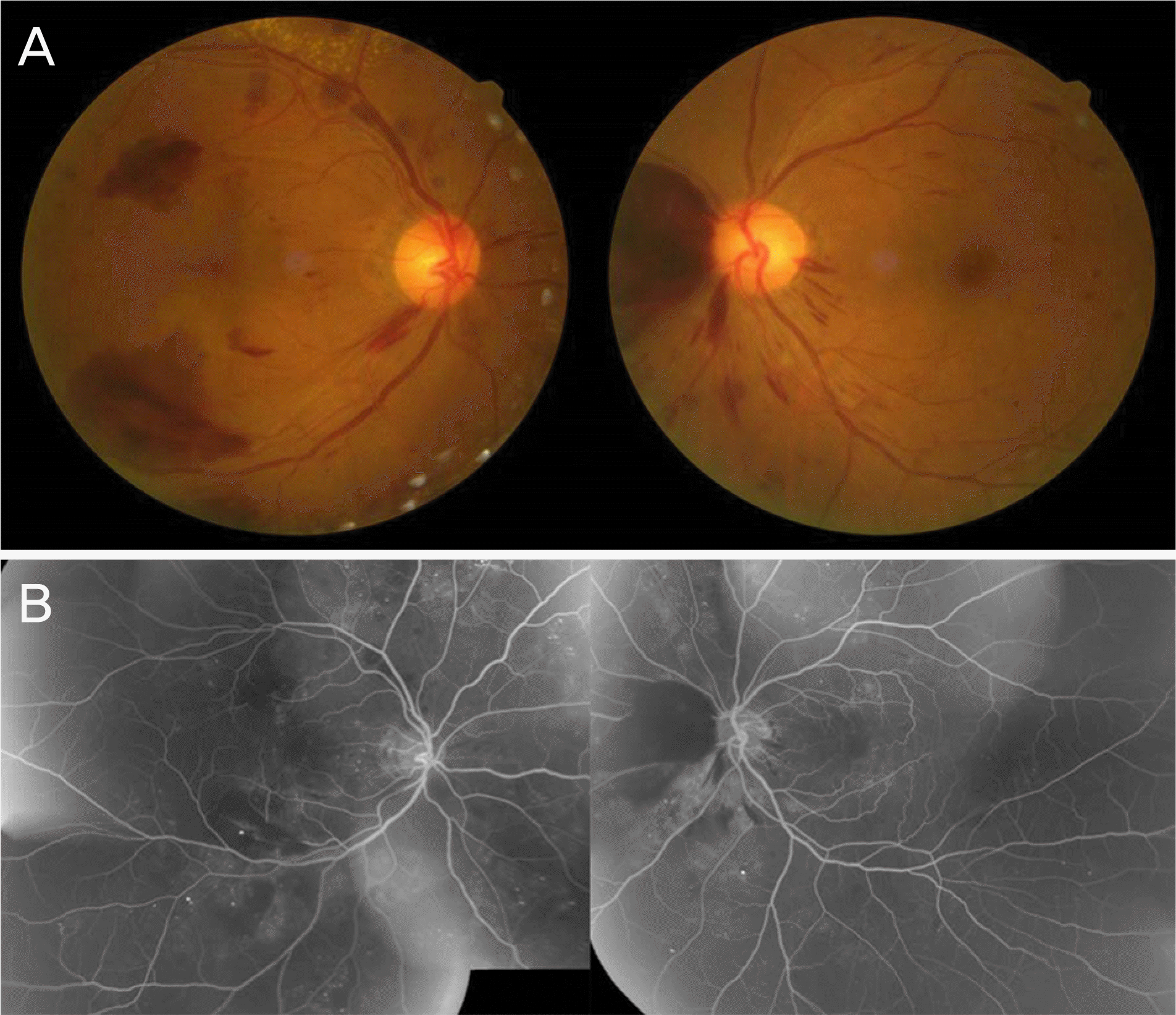
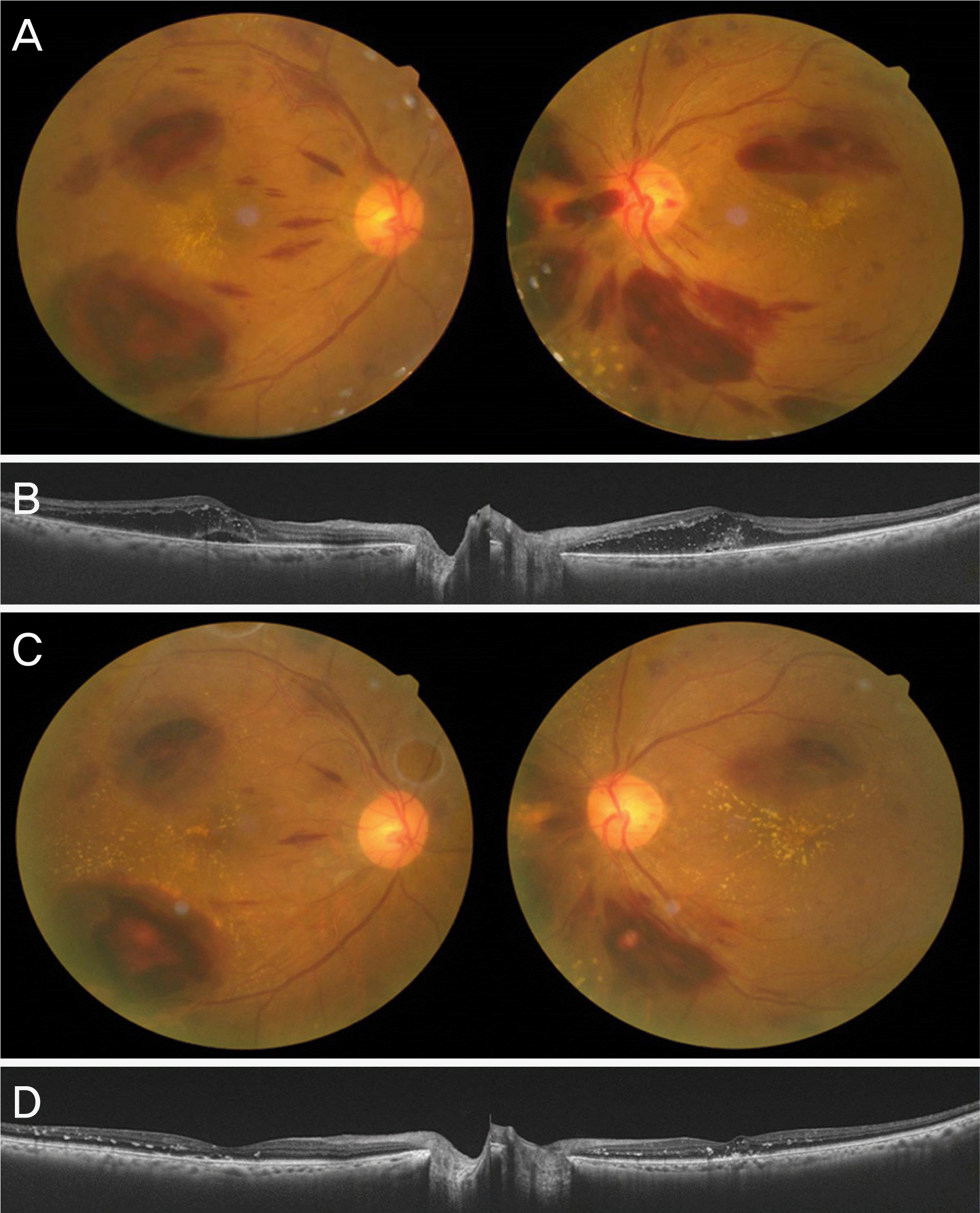
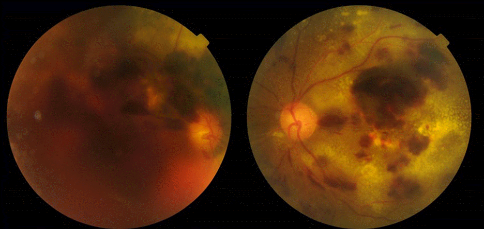
A 52-year-old female presented with a complaint of decreased visual acuity, which was 0.63 in both eyes. The patient had received a splenectomy, was receiving systemic treatment for chronic ITP, and had a history of diabetes mellitus and hypertension. Multiple retinal and subretinal hemorrhages and Roth spots were found on fundus examination. Fluorescein angiography revealed microaneurysms and hemorrhages in all four quadrants of the retina. The patient's platelet count was 38,000/pL. The patient was diagnosed with non-proliferative diabetic retinopathy and ITP-associated retinopathy, and underwent panretinal photocoagulation. Sudden visual loss was noted 4 months later, at which time the patient's visual acuity was 0.1 in both eyes, and her platelet count was 7,000/gL For 2 years, the patient's platelet count was not controlled, and remained between 12,000–19,000/pL despite active medical treatment. Macular edema did not improve with intravitreal triamcinolone, dexamethasone, and bevacizumab. Retinal hemorrhages were not absorbed and visual acuity decreased to light perception in the right eye and counting fingers in the left eye.
REFERENCES
1). Sodhi PK, Jose R. Subconjunctival hemorrhage: the first presenting clinical feature of idiopathic thrombocytopenic purpura. Jpn J Ophthalmol. 2003; 47:316–8.

2). Goel N, Arora S, Jain P, Ghosh B. Massive subretinal and vitreous hemorrhages at presentation in idiopathic thrombocytopenic purpura: report of a case and review of literature. Clin Exp Optom. 2014; 97:270–3.
3). Gruson B, Bussel JB. Immune Thrombocytopenia. Rose NR, Mackay IR, editors. The Autoimmune Diseases. 5th. London: Elsevier Academic Press;2013. chap. 47.

4). Rubenstein RA, Yanoff M, Albert DM. Thrombocytopenia, anemia and retinal hemorrhage. Am J Ophthalmol. 1968; 65:435–9.

5). Meyer CH, Callizo J, Mennel S, Schmidt JC. Complete resorption of retinal hemorrhages in idiopathic thrombocytopenic purpura. Eur J Ophthalmol. 2007; 17:128–9.

6). Okuda A, Inoue M, Shinoda K, Tsubota K. Massive bilateral vitreoretinal hemorrhage in patient with chronic refractory idiopathic thrombocytopenic purpura. Graefes Arch Clin Exp Ophthalmol. 2005; 243:1190–3.

7). Majji AB, Bhatia K, Mathai A. Spontaneous bilateral peripapillary, subhyaloid and vitreous hemorrhage with severe anemia secondary to idiopathic thrombocytopenic purpura. Indian J Ophthalmol. 2010; 58:234–6.

8). Ling R, James B. White-centred retinal haemorrhages (Roth spots). Postgrad Med J. 1998; 74:581–2.

9). Ciardella AP, Klancnik J, Schiff W, et al. Intravitreal triamcinolone for the treatment of refractory diabetic macular oedema with hard exudates: an optical coherence tomography study. Br J Ophthalmol. 2004; 88:1131–6.

10). Larsson J, Kifley A, Zhu M, et al. Rapid reduction of hard exudates in eyes with diabetic retinopathy after intravitreal triamcinolone: data from a randomized, placebo-controlled, clinical trial. Acta Ophthalmol. 2009; 87:275–80.

11). Cakir M, Kapran Z, Basar D, et al. Optical coherence tomography evaluation of macular edema after intravitreal triamcinolone acetonide in patients with parafoveal telangiectasis. Eur J Ophthalmol. 2006; 16:711–7.

12). Jarin RR, Teoh SC, Lim TH. Resolution of severe macular oedema in adult Coat's syndrome with high-dose intravitreal triamcinolone acetonide. Eye (Lond). 2006; 20:163–5.

13). Kocak N, Saatci AO, Arikan G, Bajin FM. Combination of photodynamic therapy, intravitreal triamcinolone injection, and standard laser photocoagulation in radiation retinopathy: a case report. Ann Ophthalmol (Skokie). 2006; 38:243–7.
14). Saatci AO, Doruk HC, Yaman A. Intravitreal dexamethasone implant (ozurdex) in coats' disease. Case Rep Ophthalmol. 2013; 4:122–8.

15). Mehta H, Fraser-Bell S, Yeung A, et al. Efficacy of dexamethasone versus bevacizumab on regression of hard exudates in diabetic maculopathy: data from the BEVORDEX randomized clinical trial. Br J Ophthalmol. 2016; 100:1000–4.
16). Stringa F, Marzi F, Gianni L, et al. Long-term follow-up of anatomical and functional macular changes after a single intravitreal implant of dexamethasone 0.7 mg for radiation macular edema secondary to proton beam therapy for choroidal melanoma. Int Med Case Rep J. 2016; 9:377–83. eCollection 2016.

Figure 1.
Fundus photograph and fluorescein angiography at the initial visit. (A) Fundus photograph at the initial visit shows multiple retinal and subretinal hemorrhages not involving macula. (B) Fluorescein angiography shows microaneurysms at four quadrants of each eye.

Figure 2.
Fundus photograph and optical coherence tomography after panretinal photocoagulation and intravitreal triamcinolone injection. (A) Fundus photograph at 4 months after panretinal photocoagulation. Retinal and subretinal hemorrhages involving macula and macular hard exudates are noted. (B) Optical coherence tomography shows intra- and sub-retinal fluid and reflective dots in the inner retina. (C) Fundus photograph 1 month after intravitreal triamcinolone injection. Retinal and subretinal hemorrhages are decreased. (D) Optical coherence tomography shows decreased intra- and sub-retinal fluid and foveal pit has been recovered.


 PDF
PDF ePub
ePub Citation
 Print
Print


 XML Download