Abstract
Purpose
We report two cases of retinal folds developing after pars plana vitrectomy in patients exhibiting rhegmatogenous retinal detachment.
Case summary
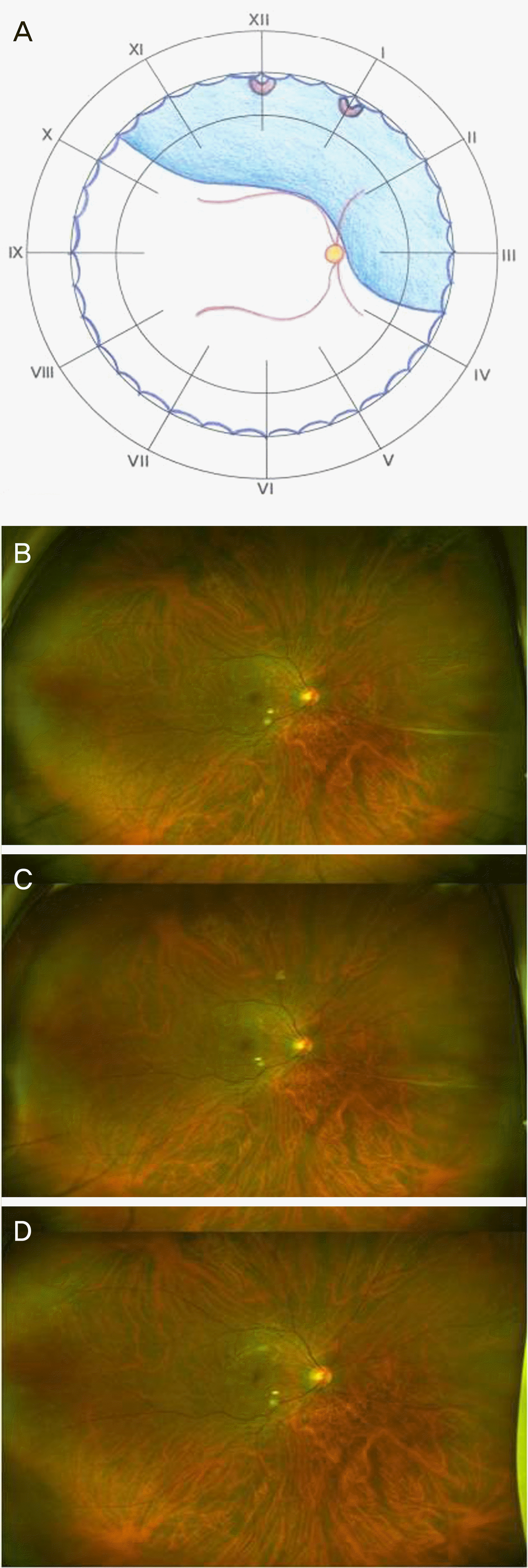
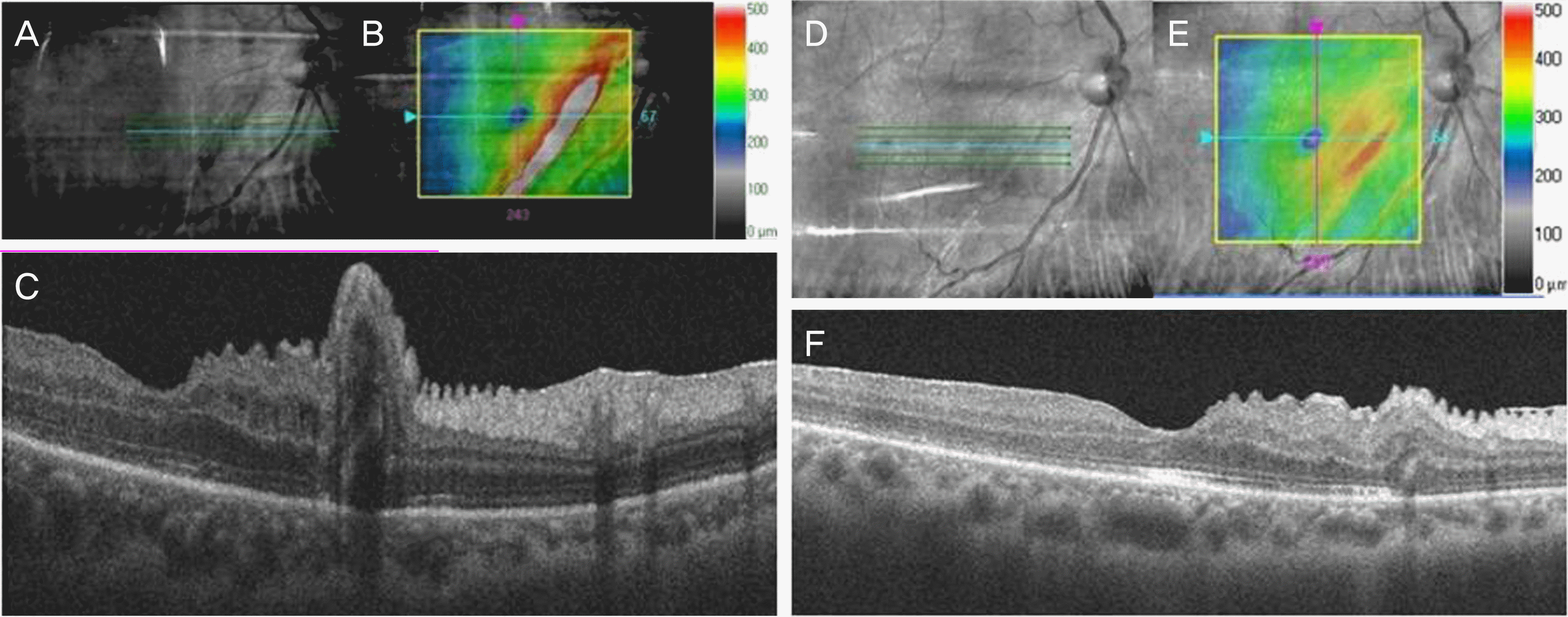
(Case 1) A healthy 52-year-old male visited our clinic complaining of blurred vision in his right eye. His visual acuity was 0.8 in that eye. Fundal examinations revealed upper retinal detachment and retinal tears at the 12 and 1 o'clock positions. He underwent pars plana vitrectomy with gas injection, and 1 week later, the retina was reattached. A retinal fold was detected at the 4 o'clock position; the fold extended for two disc diameters from the optic disc to the equator. The fold resolved spontaneously after 3 months. (Case 2) A 59-year-old male visited our clinic complaining of blurred vision in his right eye. His visual acuity was “counting fingers” in that eye. Fundal examination revealed a retinal tear at the 11 o'clock position and upper retinal detachment involving the macula. He underwent pars plana vitrectomy with gas injection. A retinal fold was detected in the temporal region of the disc running from the 7 o'clock position to the equator. Over 11 months of observation without treatment, optical coherence tomography (OCT) revealed that the retinal fold resolved.
Go to : 
REFERENCES
1). dell'Omo R, Costagliola C. Longitudinal study of macular folds by spectral-domain optical coherence tomography. Am J Ophthalmol. 2012; 154:757–8. author reply 8-9.
2). Iafe NA, Law S, Sarraf D, Tsui I. Outer retinal folds following pars plana vitrectomy with membrane peel. Retin Cases Brief Rep. 2017; 11(Suppl 1):S31–3.

3). Pavan PR. Retinal fold in macula following intraocular gas. An avoidable complication of retinal detachment surgery. Arch Ophthalmol. 1984; 102:83–4.
4). El-Amir AN, Every S, Patel CK. Repair of macular fold following retinal reattachment surgery. Clin Exp Ophthalmol. 2007; 35:791–2.

5). Ruiz-Moreno JM, Montero JA. Sliding macular fold following retinal detachment surgery. Graefes Arch Clin Exp Ophthalmol. 2011; 249:301–3.

6). Gotzaridis E, Georgalas I, Ladas I. Spontaneous regression of a retinal fold a year after scleral buckling and intravitreal injection of gas. Eye (Lond). 2010; 24:394–6.

7). Song YJ, Kim YB, Jo YJ. Management of macular folds using submacular BSS injection and partial fluid-gas exchange. J Korean Ophthalmol Soc. 2003; 44:2022–7.
8). Albert DM, Jakobiec FA. Principles and Practice of Ophthalmology. 2nd. Philadelphia: W.B. Saunders Co.;2000. p. 889–98.
9). Heinrich H, Silvia B. Retinal folds following retinal detachment surery. Ophthalmologia. 2011; 226(Suppl 1):18–26.
10). Hayashi A, Usui S, Kawaguchi K, et al. Retinal changes after retinal translocation surgery with scleral imbrication in dog eyes. Invest Ophthalmol Vis Sci. 2000; 41:4288–92.
11). Trinh L, Glacet-Bernard A, Colasse-Marthelot V, et al. Macular fold following retinal detachment surgery. J Fr Ophtalmol. 2006; 29:995–9.
Go to : 
 | Figure 1.Fundus photography of case 1 before and after vitrectomy. (A) Upper retinal detachment was detected with retinal tear (at 12 and 1 o'clock) before vitrectomy. (B) Retinal fold was detected from the optic disc to the equator at 3 weeks after vitrectomy. (C) Retinal fold remained at 2 months after vitrectomy. (D) Retinal fold was relieved spontaneously after 3 months. |
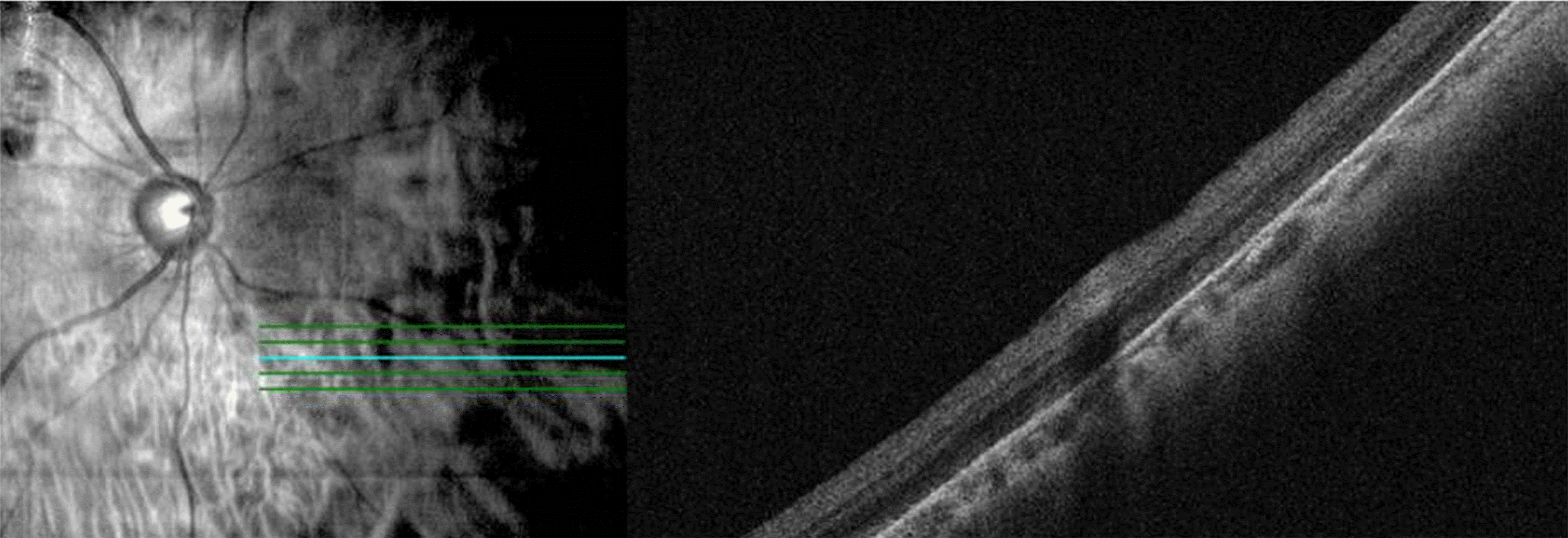
 | Figure 2.Optical coherence tomography findings of case 1 at 3 months after vitrectomy. Retinal fold was not detected, but hyperreflective lesions were detected at inner retinal layer. |
 | Figure 3.Serial fundus photography of case 2 before and after vitrectomy. (A) Upper retinal detachment involving the macular was detected with retinal tear (at 11 o'clock) before vitrectomy. (B) Retinal fold was detected from mid periphery to the equator at 10 days after vitrectomy. (C) Retinal fold was partial relieved at 11 months after vitrectomy. |
 | Figure 4.Consecutive spectral-domain optical coherence tomography (OCT) findings of case 2 at 10 days after vitrectomy (A-C) and at 11 months after vitrectomy (D-F). (A) Infrared fundus image. Blue line: level of OCT cut. (B) Fundus image with cube scan overlay showed the range of retinal fold. (C) Raster scan. Retinal fold was detected through full thickness retinal layer with nerve fiber layer undulation and inner segment ellipsoid zone disruption at 10 days after vitrectomy. (D) Infrared fundus image. Blue line: level of OCT image. (E) Fundus image with cube scan overlay. The range of retinal folds was reduced in macular cube scan. (F) Retinal fold was relieved at 11 months after vitrectomy. Retinal fold's thickness was decreased and inner segment ellipsoid zone disruption was ameliorated in Raster scan image. The range of retinal folds was reduced in macular cube scan image. |


 PDF
PDF ePub
ePub Citation
 Print
Print


 XML Download