

 PDF
PDF ePub
ePub Citation
 Print
Print


Abstract
Purpose
To assess the effects of structural changes in the lamina cribrosa (LC) and the status of the autonomic nervous system on disc hemorrhages (DHs).
Methods
A retrospective study was performed on 68 eyes of 68 patients with primary open-angle glaucoma and optic DHs. We divided the patients into two groups using optical coherence tomography according to the presence of LC defects, and then compared both groups. We also analyzed autonomic nervous system function using the heart rate variability test, and compared the two groups.
Results
Eyes with LC defects had significantly longer axial lengths than those without defects (p = 0.029), and the DH was located more proximally (p < 0.001). A significantly larger proportion of eyes without LC defects had configurational optic disc changes such as optic disc rim notching, focal rim thinning, or generalized thinning (p = 0.001). On heart rate variability testing, the group without LC defects had a significantly higher “low frequency/high frequency ratio” than the group with defects (p = 0.008).
Conclusions
There was a difference in the clinical features of DH between eyes with and without LC defects. Eyes with LC defects were more myopic and the proximal part of the DH tended to be on the disc cup or characterized by peripapillary atrophy. These results suggest that the DH developed due to a mechanical cause in eyes with LC defects. Patients without LC defects had a more dysregulated autonomic nervous system. The DH location was related to disc rim notching and neural rim losses, which implies ischemia as the pathogenic mechanism involved in the development of DH in eyes without LC defects. Therefore, more careful observations of the LC would facilitate a better understanding of the specific pathogenic mechanisms underlying DH.
Figures and Tables
Table 1
Characteristics of open-angle glaucoma patients with single or recurrent disc hemorrhage during follow-up periods
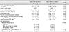
Table 2
Characteristics of open-angle glaucoma patients with single or recurrent disc hemorrhage during follow-up periods


References
1. Quigley HA, Addicks EM, Green WR, Maumenee AE. Optic nerve damage in human glaucoma. II. The site of injury and susceptibility to damage. Arch Ophthalmol. 1981; 99:635–649.
2. Grieshaber MC, Terhorst T, Flammer J. The pathogenesis of optic disc splinter haemorrhages: a new hypothesis. Acta Ophthalmol Scand. 2006; 84:62–68.

3. Sugiyama K, Tomita G, Kitazawa Y, et al. The associations of optic disc hemorrhage with retinal nerve fiber layer defect and peripapillary atrophy in normal-tension glaucoma. Ophthalmology. 1997; 104:1926–1933.
4. Morrison JC, Jerdan JA, Dorman ME, Quigley HA. Structural proteins of the neonatal and adult lamina cribrosa. Arch Ophthalmol. 1989; 107:1220–1224.
5. Morrison JC, L'Hernault NL, Jerdan JA, Quigley HA. Ultrastructural location of extracellular matrix components in the optic nerve head. Arch Ophthalmol. 1989; 107:123–129.
6. Burgoyne CF. A biomechanical paradigm for axonal insult within the optic nerve head in aging and glaucoma. Exp Eye Res. 2011; 93:120–132.
7. Park SC, Hsu AT, Su D, et al. Factors associated with focal lamina cribrosa defects in glaucoma. Invest Ophthalmol Vis Sci. 2013; 54:8401–8407.
8. Kiumehr S, Park SC, Syril D, et al. In vivo evaluation of focal lamina cribrosa defects in glaucoma. Arch Ophthalmol. 2012; 130:552–559.
9. You JY, Park SC, Su D, et al. Focal lamina cribrosa defects associated with glaucomatous rim thinning and acquired pits. JAMA Ophthalmol. 2013; 131:314–320.
10. Tatham AJ, Miki A, Weinreb RN, et al. Defects of the lamina cribrosa in eyes with localized retinal nerve fiber layer loss. Ophthalmology. 2014; 121:110–118.
11. Brown CM, Dutsch M, Michelson G, et al. Impaired cardiovascular responses to baroreflex stimulation in open-angle and normal-pressure glaucoma. Clin Sci (Lond). 2002; 102:623–630.
12. Clark CV, Mapstone R. Systemic autonomic neuropathy in open-angle glaucoma. Doc Ophthalmol. 1986; 64:179–185.
13. Kashiwagi K, Tsumura T, Ishii H, et al. Circadian rhythm of autonomic nervous function in patients with normal-tension glaucoma compared with normal subjects using ambulatory electrocardiography. J Glaucoma. 2000; 9:239–246.
14. Nicolela MT, Drance SM. Various glaucomatous optic nerve appearances: clinical correlations. Ophthalmology. 1996; 103:640–649.
15. Park HY, Hwang YS, Park CK. Ocular characteristics associated with the location of focal lamina cribrosa defects in open-angle glaucoma patients. Eye (Lond). 2017; 31:578–587.
16. Choi BM, Noh GJ. Heart rate variability. Intraven Anesth. 2004; 8:45–86.
17. Drance SM. Disc hemorrhages in the glaucomas. Surv Ophthalmol. 1989; 33:331–337.
18. Sonnsjö B, Dokmo Y, Krakau T. Disc haemorrhages, precursors of open angle glaucoma. Prog Retin Eye Res. 2002; 21:35–56.
19. Budenz DL, Anderson DR, Feuer WJ, et al. Detection and prognostic significance of optic disc hemorrhages during the Ocular Hypertension Treatment Study. Ophthalmology. 2006; 113:2137–2143.
20. Bengtsson B, Leske MC, Yang Z, et al. Disc hemorrhages and treatment in the early manifest glaucoma trial. Ophthalmology. 2008; 115:2044–2048.
21. Drance S, Anderson DR, Schulzer M;. Risk factors for progression of visual field abnormalities in normal-tension glaucoma. Am J Ophthalmol. 2001; 131:699–708.
22. Jonas JB, Martus P, Budde WM, Hayler J. Morphologic predictive factors for development of optic disc hemorrhages in glaucoma. Invest Ophthalmol Vis Sci. 2002; 43:2956–2961.
23. Ahn JK, Park KH. Morphometric change analysis of the optic nerve head in unilateral disk hemorrhage cases. Am J Ophthalmol. 2002; 134:920–922.
24. Nitta K, Sugiyama K, Higashide T, et al. Does the enlargement of retinal nerve fiber layer defects relate to disc hemorrhage or progressive visual field loss in normal-tension glaucoma? J Glaucoma. 2011; 20:189–195.
25. Choi J, Kim KH, Jeong J, et al. Circadian fluctuation of mean ocular perfusion pressure is a consistent risk factor for normal-tension glaucoma. Invest Ophthalmol Vis Sci. 2007; 48:104–111.
26. Delaney Y, Walshe TE, O'Brien C. Vasospasm in glaucoma: clinical and laboratory aspects. Optom Vis Sci. 2006; 83:406–414.
27. Flammer J, Orgül S, Costa VP, et al. The impact of ocular blood flow in glaucoma. Prog Retin Eye Res. 2002; 21:359–393.
28. Sugiyama T, Utsunomiya K, Ota H, et al. Comparative study of cerebral blood flow in patients with normal-tension glaucoma and control subjects. Am J Ophthalmol. 2006; 141:394–396.
29. Usui T, Iwata K, Shirakashi M, Abe H. Prevalence of migraine in low-tension glaucoma and primary open-angle glaucoma in Japanese. Br J Ophthalmol. 1991; 75:224–226.
30. Park HY, Jeong HJ, Kim YH, Park CK. Optic disc hemorrhage is related to various hemodynamic findings by disc angiography. PLoS One. 2015; 10:e0120000. DOI: 10.1371/journal.pone.0120000. eCollection 2015.
31. Park HY, Jung KI, Na KS, et al. Visual field characteristics in normal-tension glaucoma patients with autonomic dysfunction and abnormal peripheral microcirculation. Am J Ophthalmol. 2012; 154:466–475. e1.
32. Pagani M, Lombardi F, Guzzetti S, et al. Power spectral density of heart rate variability as an index of sympatho-vagal interaction in normal and hypertensive subjects. J Hypertens Suppl. 1984; 2:S383–S385.
33. Pagani M, Lombardi F, Guzzetti S, et al. Power spectral analysis of heart rate and arterial pressure variabilities as a marker of sympatho-vagal interaction in man and conscious dog. Circ Res. 1986; 59:178–193.
34. Malliani A, Pagani M, Lombardi F, Cerutti S. Cardiovascular neural regulation explored in the frequency domain. Circulation. 1991; 84:482–492.
35. Grassi G. Assessment of sympathetic cardiovascular drive in human hypertension: achievements and perspectives. Hypertension. 2009; 54:690–697.
 XML Download