

 PDF
PDF ePub
ePub Citation
 Print
Print


Abstract
Purpose
To define risk factors for and to analyze changes in hyperopic refractive error during development of postoperative exotropia (XT) after bilateral medial rectus (BMR) recession to treat infantile esotropia.
Methods
We retrospectively examined 50 patients with infantile esotropia who underwent BMR recession from January 2005 to December 2010. All were <10 years of age and underwent S36 months of follow-up. We recorded age at operation, the preoperative strabismus angle, the extent of medial rectus recession, strabismus status, pre- and post-operative changes in the refractive errors of both eyes, any postoperative overcorrection, any dissociated vertical deviation (DVD), and inferior oblique overaction (IOOA) status.
Results
Consecutive XT developed in 18 (36%) patients. The preoperative refractive error was +0.90 ± 0.79 D in the consecutive XT group and +1.94 ± 1.48 D in the surgical success (SS) group (p = 0.019). The extent of hyperopic decrease was significantly greater in the consecutive XT group than the SS group (consecutive XT group: 1.59 ± 1.38 D, SS group: 2.86 ± 1.97 D) at 3 years of post-operative follow-up (p = 0.008). Postoperative IOOA was detected in 10 (70.5%) patients in the consecutive XT group and 3 (29.55%) in the SS group (p = 0.002). No significant between-group difference in the incidence of overcorrection or DVD was apparent.
Go to : 
REFERENCES
1). Helveston EM, Neely DF, Stidham DB, et al. Results of early alignment of congenital esotropia. Ophthalmology. 1999; 109:1716–26.
2). Beneish R, Williams F, Polomeno RC, Little JM. Consecutive exotropia after correction of hyperopia. Can J Opthalmol. 1981; 16:16–8.
3). Kim MJ, Jin YH. Spontaneous consecutive exotropia. J Korean Ophthalmol Soc. 1999; 40:2918–22.
4). Swan KC. Accommodative esotropia long range follow-up. Ophthalmology. 1983; 90:1141–5.

5). Weir CR, Cleary M, Dutton GN. Spontaneous consecutive exotropia in children with motor fusion. Br J Ophthalmol. 2001; 85:242–3.
6). Bradbury JA, Doran RM. Secondary exotropia: a retrospective analysis of matched cases. J Pediatr Ophthalmol Strabismus. 1993; 30:163–6.
7). Yazawa K. Postoperative exotropia. J Pediatr Ophthalmol Strabismus. 1981; 18:58–64.
8). Donaldson MJ, Forrest MP, Gole GA. The surgical management of consecutive exotropia. J AAPOS. 2004; 8:230–6.
9). Patel AS, Simon JW, Lininger LL. Bilateral lateral rectus recession for consecutive exotropia. J AAPOS. 2000; 4:291–4.
10). Sachdeva V, Mittal V, Reddy PN, et al. Dose-effect relationship of medial rectus muscle advancement for consecutive exotropia. J AAPOS. 2012; 16:314. author reply 314-5.
11). Chatzistefanou KI, Droutsas KD, Chimonidou E. Reversal of unilateral medial rectus recession and lateral rectus resection for the correction of consecutive exotropia. Br J Ophthalmol. 2009; 93:742–6.
12). Mohan K, Sharma A, Pandav SS. Unilateral lateral rectus muscle recession and medial rectus muscle resection with or without advancement for postoperative consecutive exotropia. J AAPOS. 2006; 10:220–4.
13). Folk ER, Miller MT, Chapman L. Consecutive exotropia following surgery. Br J Ophthalmol. 1983; 67:546–8.
14). Stager DR, Weakley DR Jr, Everett M, Birch EE. Delayed consecutive exotropia following 7-millimeter bilateral medial rectus recession for congenital esotropia. J Pediatr Ophthalmol Strabismus. 1994; 31:147–50. discussion 151-2.
15). Oggz V, Arvas S, Yolar M, et al. Consecutive exotropia following strabismus surgery. Ophthalmologica. 2002; 216:246–8.
16). Knapp P. The surgical treatment of persistent horizontal strabismus. Trans Am Ophthalmol Soc. 1965; 63:75–90.
17). Ohtsuki H, Hasebe S, Tadokoro Y, et al. Advancement of medial rectus muscle to the original insertion for consecutive exotropia. J Pediatr Ophthalmol Strabismus. 1993; 30:301–5.
18). Pickering JD, Simon JW, Lininger LL, et al. Exaggerated effect of bilateral medial rectus recession in developmentally delayed children. J Pediatr Ophthalmol Strabismus. 1994; 31:374–7.
19). Lee JR, Roh YB. The factors affecting consecutive exotropia with angle of 20 prism diopters or more following surgery for esotropia. J Korean Ophthalmol Soc. 1995; 36:1778–83.
20). Berk AT, Kofak N, Ellidokuz H. Treatment outcomes in refractive accommodative esotropia. J AAPOS. 2004; 8:384–8.
21). Tang SM, Chan RY, Bin Lin S, et al. Refractive errors and concomitant strabismus: a systematic review and meta-analysis. Sci Rep. 2016; 6:35177.
22). Kim HK, Chung HJ, Park SH, Shin SY. Consecutive exotropia after bilateral medial rectus recession for infantile esotropia. J Korean Ophthalmol Soc. 2009; 50:1712–6.
23). Louwagie CR, Diehl NN, Greenberg AE, Mohney BG. Long-term follow-up of congenital esotropia in a population-based cohort. J AAPOS. 2009; 13:8–12.
24). Bae SH, Choi DG. Clinical features and surgical outcomes of infantile esotropia according to the age at surgery. J Korean Ophthalmol Soc. 2008; 49:1961–7.
Go to :
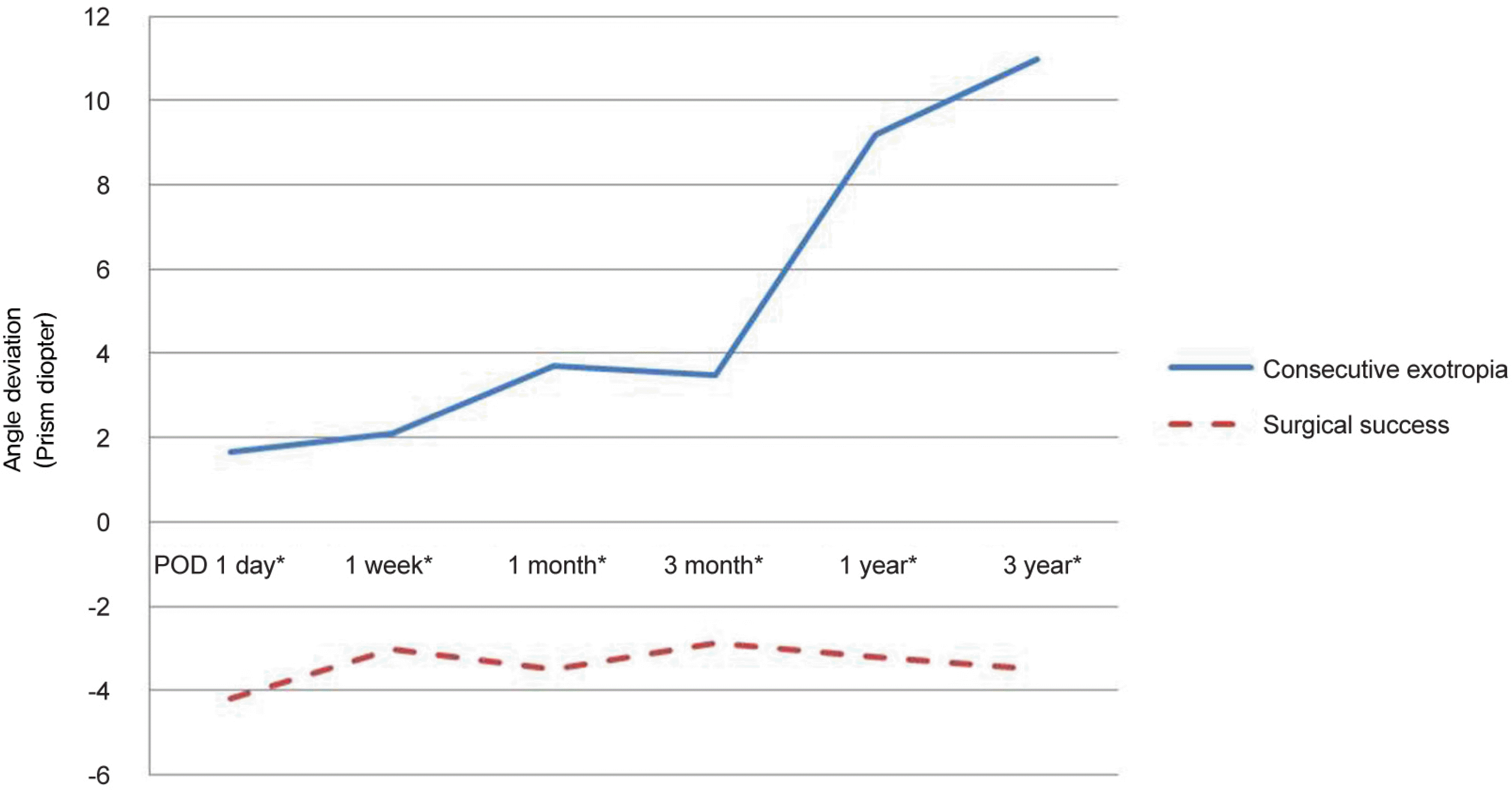 | Figure 1.Postoperative angle of deviation according to postoperative durations between consecutive exotropia (XT) group and surgical success group. The amount of postoperative exotropic shift was higher in the consecutive exotropia group. POD = postoperative day. *Statistically significant difference (Mann-Whitney U-test: p < 0.05). |
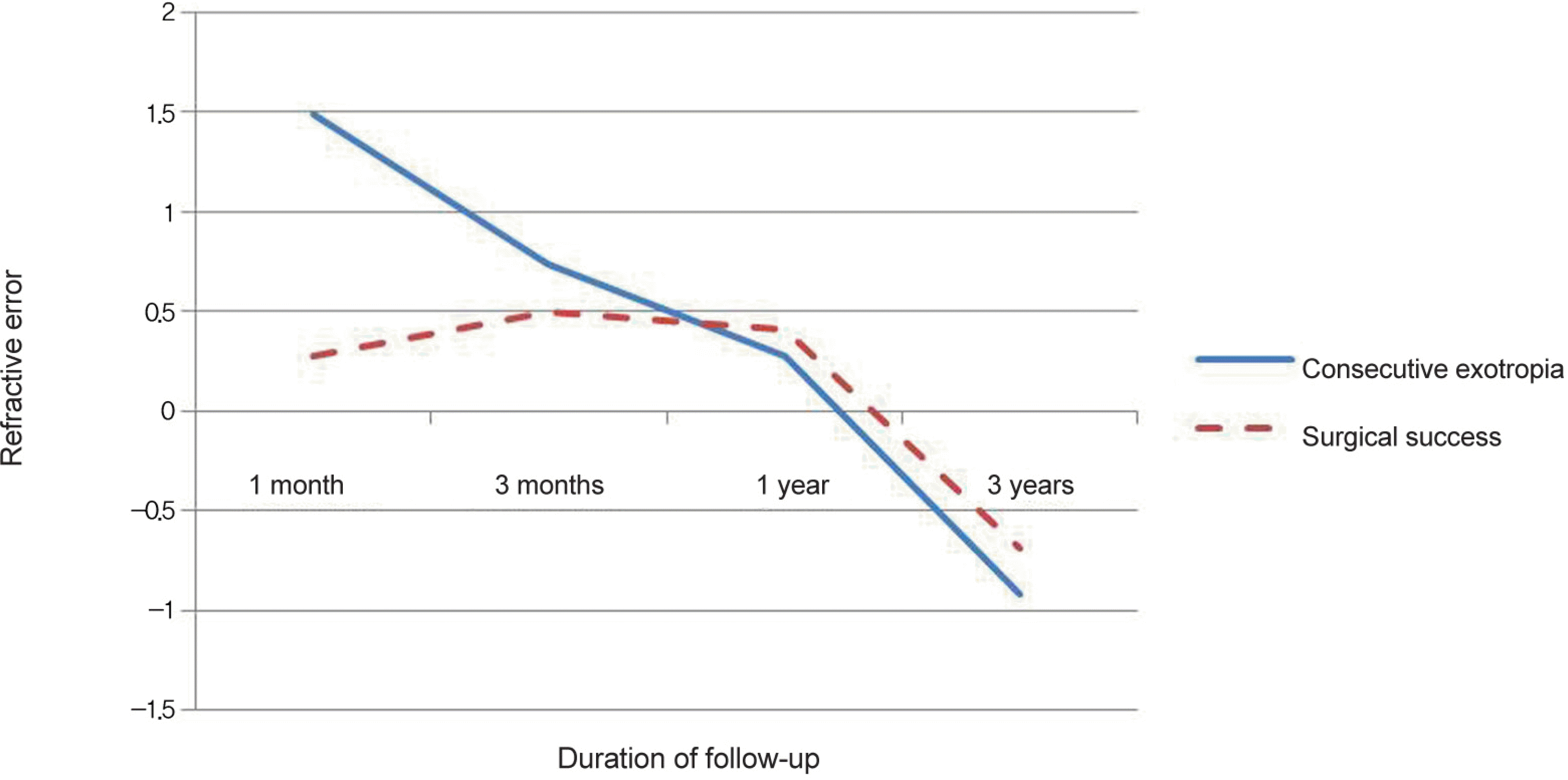 | Figure 2.Changes of hyperopic refractive error during the follow-up after surgery for postoperative refractive error at 1 month, 3 months, 1 year, and 3 years postoperatively in each group. The extent of hyperopic decrease was significantly greater in the consecutive exotropia (XT) group than the surgical success (SS) group. *Statistically significant difference (Mann-Whitney U-test: p < 0.05). |
Table 1.
Patient demographics, preoperative data, operative and postoperative data
| Total | Consecutive exotropia | Surgical success | p-value | |
|---|---|---|---|---|
| No. patients | 44 | 18 | 26 | |
| Preoperative | ||||
| Ratio (female:male) | 26:18 | 11:15 | 10:8 | 0.358† |
| Mean age at surgery (years) | 3.92 ± 1.59 | 3.66 ± 1.87 | 4.10 ± 1.38 | 0.429* |
| Mean refractive errors of both eyes (D) | 1.32 ± 1.23 | 1.94 ± 1.48 | 0.90 ± 0.79 | 0.019* |
| Preoperative deviation (PD) | ||||
| At distance | 34.26 ± 9.96 | 33.89 ± 11.45 | 34.57 ± 9.03 | 0.689* |
| Near | 35.20 ± 12.14 | 35.83 ± 15.07 | 34.77 ± 9.93 | 0.874* |
| IOOA (n, %) | 7 (15.9) | 5 (28.8) | 2 (7.7) | 0.103† |
| DVD (n, %) | 2 (4.5) | 2 (11.1) | 0 (0) | 0.162† |
| Postoperative | ||||
| Medial rectus recess amount (mm) | 4.69 ± 0.75 | 4.43 ± 0.93 | 4.87 ± 0.54 | 0.145* |
| Amount of hyperopic decrease (PD/3yr) | 2.62 ± 0.74 | 2.56 ± 0.75 | 2.66 ± 0.75 | 0.008* |
| Overcorrection (n, %) | 0 (0) | 0 (0) | 0 (0) | |
| IOOA (n, %) | 13 (29.5) | 10 (70.5) | 3 (11.5) | 0.003† |
| DVD (n, %) | 2 (4.5) | 2(11.11) | 0 (0) | 0.162† |
| Follow-up (months) | 49.36 ± 17.16 | 53.44 ± 25.26 | 49.36 ± 17.16 | 0.145* |
| Mean age at last follow up | 7.66 ± 2.09 | 7.26 ± 2.47 | 7.94 ± 1.77 | 0.518* |
Table 2.
Postoperative refractive error at 1 month, 3 months, 1 year, and 3 years postoperatively in each group
| Time after surgery |
Refractive error |
||
|---|---|---|---|
| Consecutive exotropia | Surgical success | p-value | |
| 1 month | 1.49 ± 1.28 | 0.28 ± 0.65 | <0.001* |
| 3 months | 0.74 ± 0.96 | 0.45 ± 0.70 | 0.243* |
| 1 year | 0.28 ± 0.96 | 0.41 ± 0.63 | 0.724* |
| 3 years | −0.92 ± 1.49 | −0.69 ± 1.07 | 0.151* |
 XML Download