Abstract
Purpose
To evaluate the effect of periosteal fixation in patients with large-angle paralytic strabismus that was not corrected through conventional strabismus surgery.
Methods
Four eyes of three patients with large-angle paralytic strabismus who underwent periosteal fixation from June 2014 to August 2014 were examined. All patients presented with exotropia > 50 prism diopters (PD). Two of them showed exotropia caused by chronic complete oculomotor nerve palsy; the other two showed exotropia caused by medial rectus muscle injury during endoscopic sinus surgery.
REFERENCES
1). Rosenbaum AL, Santiago AP. Clinical Strabismus Management: Principles and Surgical Techniques. Philadelphia: WB Saunders;1999. p. 252.
3). Lee MS, Ahn JH, Kim HY, Lee SY. Clinical study of orbital wall fracture. J Korean Ophthalmol Soc. 1997; 38:1687–93.
4). Koornneef L. Current concepts on the management of orbital blow-out fractures. Ann Plast Surg. 1982; 9:185–200.

5). Helveston EM. Surgical Management of Strabismus: An Atlas of Strabismus Surgery. 3rd. St. Louis: CV Mosby;1985. p. 254–9.
7). Morad Y, Kowal L, Scott AB. Lateral rectus muscle disinsertion and reattachment to the lateral orbital wall. Br J Ophthalmol. 2005; 89:983–5.

8). Salazar-Leon JA, Ramirez-Ortiz MA, Salas-Vargas M. The surgical correction of paralytic strabismus using fascia lata. J Pediatr Ophthalmol Strabismus. 1998; 35:27–32.

9). Srivastava KK, Sundaresh K, Vijayalakshmi P. A new surgical technique for ocular fixation in congenital third nerve palsy. J AAPOS. 2004; 8:371–7.

10). Sharma P, Gogoi M, Kedar S, Bhola R. Periosteal fixation in third-nerve palsy. J AAPOS. 2006; 10:324–7.

11). Goldberg RA, Rosenbaum AL, Tong JT. Use of apically based periosteal flaps as globe tethers in severe paretic strabismus. Arch Ophthalmol. 2000; 118:431–7.

13). Saxena R, Sinha A, Sharma P, et al. Precaruncular approach for medial orbital wall periosteal anchoring of the globe in oculomotor nerve palsy. J AAPOS. 2009; 13:578–82.

14). von Noorden GK. Binocular Vision and Ocular Motility: Theory and Management of Strabismus. 3rd. St. Louis: CV Mosby;1985. p. 369.
15). Villasenor Solares J, Riemann BI, Romanelli Zuazo AC, Riemann CD. Ocular fixation to nasal periosteum with a superior oblique tendon in patients with third nerve palsy. J Pediatr Ophthalmol Strabismus. 2000; 37:260–5.
16). Bicas HE. Asurgically implanted elastic band to restore paralyzed ocular rotations. J Pediatr Ophthalmol Strabismus. 1991; 28:10–3.
17). Collins CC, Jampolsky A, Scott AB. Artificial muscles for extraocular implantation. Invest Ophthalmol Vis Sci. 1985; 26(3 Suppl 1):80.
18). Schumacher-Feero LA, Yoo KW, Solari FM, Biglan AW. Third cranial nerve palsy in children. Am J Ophthalmol. 1999; 128:216–21.

19). Jackson E. Operations on muscles of the eye. Wiener M, Scheie HG, editors. Surgery of the eye. 3rd. New York: Grune & Stratton;1952. p. 405.
20). Saunders RA, Rogers GL. Superior oblique transposition for third nerve palsy. Ophthalmology. 1982; 89:310–6.

21). Dalman NE, Schwarcz RM, Velez FG. Suture fixation system as globe tethers in severe paralytic strabismus. J AAPOS. 2006; 10:371–2.
22). Saxena R, Sinha A, Sharma P, et al. Precaruncular periosteal anchor of medial rectus, a new technique in the management of complete external third nerve palsy. Orbit. 2006; 25:205–8.

23). Suk KW, Park JM, Kim SS, Lee SJ. Periosteal fixation in bilateral total third nerve palsy. J Korean Ophthalmol Soc. 2008; 49:1203–8.

24). Seol BR, Khwarg SI, Kim SJ. Fixation of the eyeball to the periosteum over the posterior lacrimal crest in inveterate exotropia. J Korean Ophthalmol Soc. 2014; 55:408–15.

25). Bleier BS, Schlosser RJ. Prevention and management of medial rectus injury. Otolaryngol Clin North Am. 2010; 43:801–7.

26). Kitthaweesin K, Yospaiboon Y. Dexamethasone and methyl-prednisolone in treatment of indirect traumatic optic neuropathy. J Med Assoc Thai. 2001; 84:628–34.
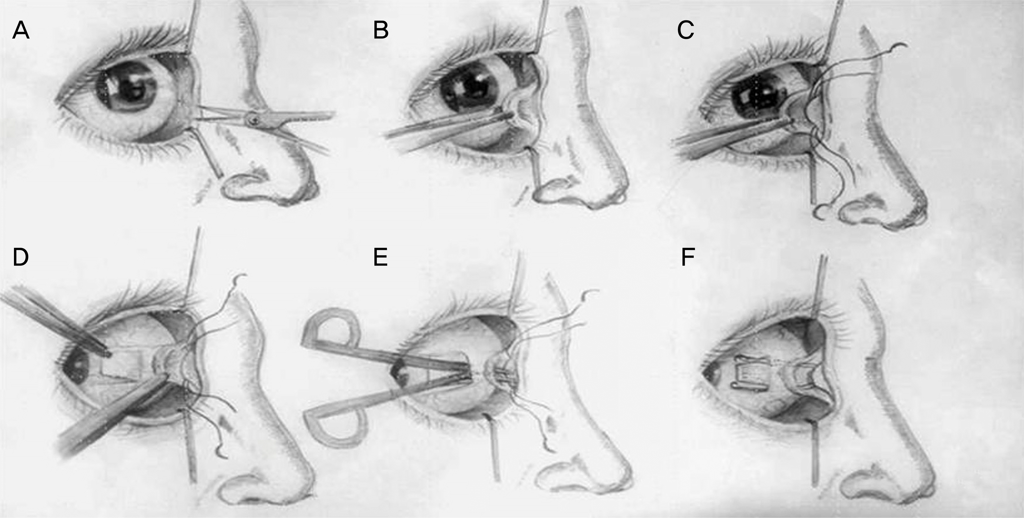
Figure 1.
Schematic drawings of periosteal fixation technique. (A) Incision is made at the precaruncular conjunctiva. (B) Blunt dissection is continued medially between caruncle and posterior lacrimal crest. (C) Nonabsorbable double armed 5-0 nylon sutures are passed through the periosteum. (D) Medial rectus insertion is exposed using limbal conjunctival approach. (E) Sutures are held with the needle tips and brought out in the sub-Tenon's space using mosquito forceps. (F) Sutures are passed on the sclera in either side of the medial rectus muscle.

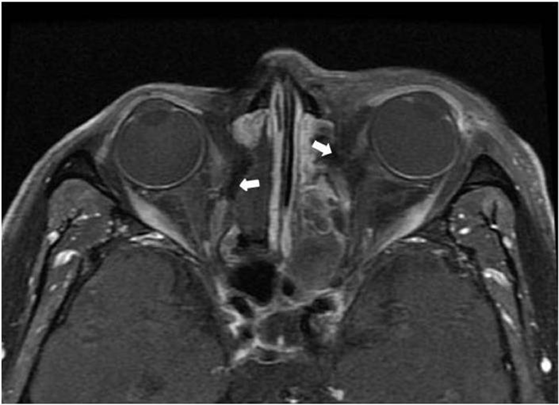
Figure 2.
Case 2. Preoperative orbital magnetic resonance imaging showing transected both medial rectus muscles (arrows).

Figure 3.
Case 2. Nine gaze photograph of a 62-year-old male patient with transected both medial rectus muscle after endoscopic sinus surgery. Before surgery, showing exotropia of above 100 prism diopters and limitation of adduction of the both eyes.

Figure 4.
Case 2. One month after periosteal fixation surgery of the left eye, showing exotropia of 40 prism diopters in primary position.

Figure 5.
Case 2. Six months after periosteal fixation of the right eye, showing exotropia of 15 prism diopters in primary position.

Table 1.
Clinical characteristics of the patients


 PDF
PDF ePub
ePub Citation
 Print
Print


 XML Download