

 PDF
PDF ePub
ePub Citation
 Print
Print


Abstract
Purpose
The aim of our study was to evaluate short-term changes in corneal surface status via Keratography in dry eye patients after placement of an absorbable collagen plug (UltraPlug™; Surgical Specialties Corporation, Reading, PA, USA).
Methods
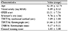
Patients (n = 20 eyes, 20 subjects) diagnosed with dry eye were recruited for this prospective, 1-month clinical trial. The lacrimal puncta were blocked using absorbable collagen plugs. We evaluated clinical parameters and symptoms 1 month later. We assessed visual acuity, the score on the Ocular Surface Disease Index (OSDI) questionnaire, and keratographic tear meniscus height (TMH) and tear break-up time (TBUT) (K5M; Oculus Optikgerate GmbH, Wetzlar, Germany). We also measured TBUT classically, the corneal fluorescein staining score, and the Schirmer test result.
Results
Significant improvements in the corneal staining and OSDI scores were observed 1 month after treatment (p = 0.006, p = 0.001, respectively), but no significant differences in the keratographic TMH and TBUT, the classical TBUT, or the Schirmer test data was observed (all p > 0.05).
Conclusions
Blocking of the lacrimal punctum with an absorbable collagen plug effectively improved clinical symptoms and objective indicators in dry eye patients, the corneal staining score and OSDI scores improved significantly. However, no significant improvements in keratographic TMH and TBUT were observed; this was of concern. Longer-term studies with more patients are required.
Figures and Tables
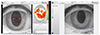 | Figure 1Examination images using Keratograph. (A) Tear break-up time (TBUT) using Keratograph. (B) Tear meniscus height (TMH) using Keratograph. From the moment the patient was last blinking, TBUT was measured by the time the ring shape of the cornea surface change and was automatically detected. TMH was measured vertically from the center of the lower eyelid border to the center of the pupil.
|

References
1. The definition and classification of dry eye disease: report of the Definition and Classification Subcommittee of the International Dry Eye WorkShop (2007). Ocul Surf. 2007; 5:75–92.
2. Friedman NJ. Impact of dry eye disease and treatment on quality of life. Curr Opin Ophthalmol. 2010; 21:310–316.

3. Miljanović B, Dana R, Sullivan DA, Schaumberg DA. Impact of dry eye syndrome on vision-related quality of life. Am J Ophthalmol. 2007; 143:409–415.
4. Marcet MM, Shtein RM, Bradley EA, et al. Safety and efficacy of lacrimal drainage system plugs for dry eye syndrome: a report by the American academy of ophthalmology. Ophthalmology. 2015; 122:1681–1687.
5. Di Pascuale MA, Goto E, Tseng SC. Sequential changes of lipid tear film after the instillation of a single drop of a new emulsion eye drop in dry eye patients. Ophthalmology. 2004; 111:783–791.
6. Baxter SA, Laibson PR. Punctal plugs in the management of dry eyes. Ocul Surf. 2004; 2:255–265.
7. Management and therapy of dry eye disease: report of the Management and Therapy Subcommittee of the International Dry Eye WorkShop (2007). Ocul Surf. 2007; 5:163–178.
8. Methodologies to diagnose and monitor dry eye disease: report of the Diagnostic Methodology Subcommittee of the International Dry Eye WorkShop (2007). Ocul Surf. 2007; 5:108–152.
9. Patel S, Murray D, McKenzie A, et al. Effects of fluorescein on tear breakup time and on tear thinning time. Am J Optom Physiol Opt. 1985; 62:188–190.
10. Mengher LS, Bron AJ, Tonge SR, Gilbert DJ. Effect of fluorescein instillation on the pre-corneal tear film stability. Curr Eye Res. 1985; 4:9–12.
11. Mengher LS, Bron AJ, Tonge SR, Gilbert DJ. A non-invasive instrument for clinical assessment of the pre-corneal tear film stability. Curr Eye Res. 1985; 4:1–7.
12. Nichols KK, Mitchell GL, Zadnik K. The repeatability of clinical measurements of dry eye. Cornea. 2004; 23:272–285.
13. Abdelfattah NS, Dastiridou A, Sadda SR, Lee OL. Noninvasive imaging of tear film dynamics in eyes with ocular surface disease. Cornea. 2015; 34:Suppl 10. S48–S52.
14. Arriola-Villalobos P, Fernandez-Vigo JI, Diaz-Valle D, et al. Assessment of lower tear meniscus measurements obtained with Keratograph and agreement with Fourier-domain optical-coherence tomography. Br J Ophthalmol. 2015; 99:1120–1125.
15. Wang X, Lu X, Yang J, et al. Evaluation of dry eye and meibomian gland dysfunction in teenagers with myopia through noninvasive keratograph. J Ophthalmol. 2016; 2016:6761206.
16. Jeong S, Lee SB. Reliability of a new non-invasive tear film break-up time measurement using a Keratograph. J Korean Ophthalmol Soc. 2016; 57:1354–1360.
17. Tian L, Qu JH, Zhang XY, Sun XG. Repeatability and reproducibility of noninvasive keratograph 5M measurements in patients with dry eye disease. J Ophthalmol. 2016; 2016:8013621.
18. McGinnigle S, Naroo SA, Eperjesi F. Evaluation of dry eye. Surv Ophthalmol. 2012; 57:293–316.
19. Koh S, Ikeda C, Watanabe S, et al. Effect of non-invasive tear stability assessment on tear meniscus height. Acta Ophthalmol. 2015; 93:e135–e139.
20. Hong J, Sun X, Wei A, et al. Assessment of tear film stability in dry eye with a newly developed keratograph. Cornea. 2013; 32:716–721.
21. Yoon KC, Im SK, Kim HG, You IC. Usefulness of double vital staining with 1% fluorescein and 1% lissamine green in patients with dry eye syndrome. Cornea. 2011; 30:972–976.
22. Bron AJ, Evans VE, Smith JA. Grading of corneal and conjunctival staining in the context of other dry eye tests. Cornea. 2003; 22:640–650.
23. Lee SJ, Kim HY, Park YM, Lee JS. Comparison of therapeutic effects of 3% diquafosol tetrasodium with aging in dry eye. J Korean Ophthalmol Soc. 2016; 57:734–741.
24. Tsubota K. Tear dynamics and dry eye. Prog Retin Eye Res. 1998; 17:565–596.
25. Jiang Y, Ye H, Xu J, Lu Y. Noninvasive Keratograph assessment of tear film break-up time and location in patients with age-related cataracts and dry eye syndrome. J Int Med Res. 2014; 42:494–502.
 XML Download