

 PDF
PDF ePub
ePub Citation
 Print
Print


Abstract
Purpose
To examine changes in meibomian glands by indirect meibography after incision and curettage of the chalazion, and to examine the relationship between meibomian gland drop out and meibomian gland dysfunction.
Methods
We performed a prospective study of 16 patients, <5 years of age, who underwent incision and curettage of the chalazion between March 2017 and June 2017. We performed indirect meibography before incision and curettage of the chalazion, and 1 week and 1 month after the procedure. Photographs of meibomian glands were rated according to their meiboscore, the break-up time (BUT) was measured, and Schirmer's test was performed.
Results
Sixteen eyes of 16 patients were included in this study. Fifteen patients visited 1 week after incision and curettage of the chalazion, and eleven patients visited at 1 month after the procedure. The mean patient age was 32.9 ± 21.9 years. There were no significant changes in the meiboscore (p = 0.092), BUT (p = 0.068), and Schirmer's test results (p = 0.972) after incision and curettage of the chalazion. In meibography, there were inflammatory changes and partial meibomian gland drop outs at the chalazion lesion before its incision and curettage. At 1 month after the procedure, only scarring remained in the chalazion lesion, and normal surrounding meibomian glands were found near the old lesions. When comparing meibography of the baseline with meibography at 1 month after incision and curettage of the chalazion, the normal area of the meibomian gland significantly increased (p = 0.041).
Conclusions
In patients with chalazions, scarring was found after incision and curettage of the lesions, and surrounding meibomian glands were well preserved when determined by indirect meibography. There was no significant correlation between meibomian gland changes after incision and curettage of the chalazion and meibomian gland dysfunction.
Go to : 
References
1. Alsuhaibani AH, Carter KD, Abràmoff MD, Nerad JA. Utility of meibography in the evaluation of meibomian glands morphology in normal and diseased eyelids. Saudi J Ophthalmol. 2011; 25:61–6.

2. Kim JH, Ro JW, Yi K, et al. Changes of the meibomian gland abdominal to age in the normal Korean population. J Korean Ophthalmol Soc. 2015; 56:13–8.
3. Finis D, Ackermann P, Pischel N, et al. Evaluation of meibomian gland dysfunction and local distribution of meibomian gland abdominal by non-contact infrared meibography. Curr Eye Res. 2015; 40:982–9.
4. Nemoto Y, Arita R, Mizota A, Sasajima Y. Differentiation between chalazion and sebaceous carcinoma by noninvasive meibography. Clin Ophthalmol. 2014; 8:1869–75.
5. Hwang HS, Park CW, Joo CK. Novel noncontact meibography with anterior segment optical coherence tomography: Hosik meibography. Cornea. 2013; 32:40–3.
6. Arita R, Itoh K, Inoue K, Amano S. Noncontact infrared abdominal to document age-related changes of the meibomian glands in a normal population. Ophthalmology. 2008; 115:911–5.
7. Aycinena AR, Achiron A, Paul M, Burgansky-Eliash Z. Incision and curettage versus steroid injection for the treatment of chalazia: a meta-analysis. Ophthal Plast Reconstr Surg. 2016; 32:220–4.
8. Arita R, Itoh K, Maeda S, et al. Proposed diagnostic criteria for abdominal meibomian gland dysfunction. Ophthalmology. 2009; 116:2058–63.e1.
9. Yokoi N, Komuro A, Yamada H, et al. A newly developed vid-eo-meibography system featuring a newly designed probe. Jpn J Ophthalmol. 2007; 51:53–6.
10. Srinivasan S, Menzies KL, Sorbara L, Jones LW. Imaging abdominal glands on a patient with chalazia in the upper and lower lids: a case report. Cont Lens Anterior Eye. 2013; 36:199–203.
11. Fukuoka S, Arita R, Shirakawa R, Morishige N. Changes in abdominal gland morphology and ocular higher-order aberrations in eyes with chalazion. Clin Ophthalmol. 2017; 11:1031–8.
Go to :
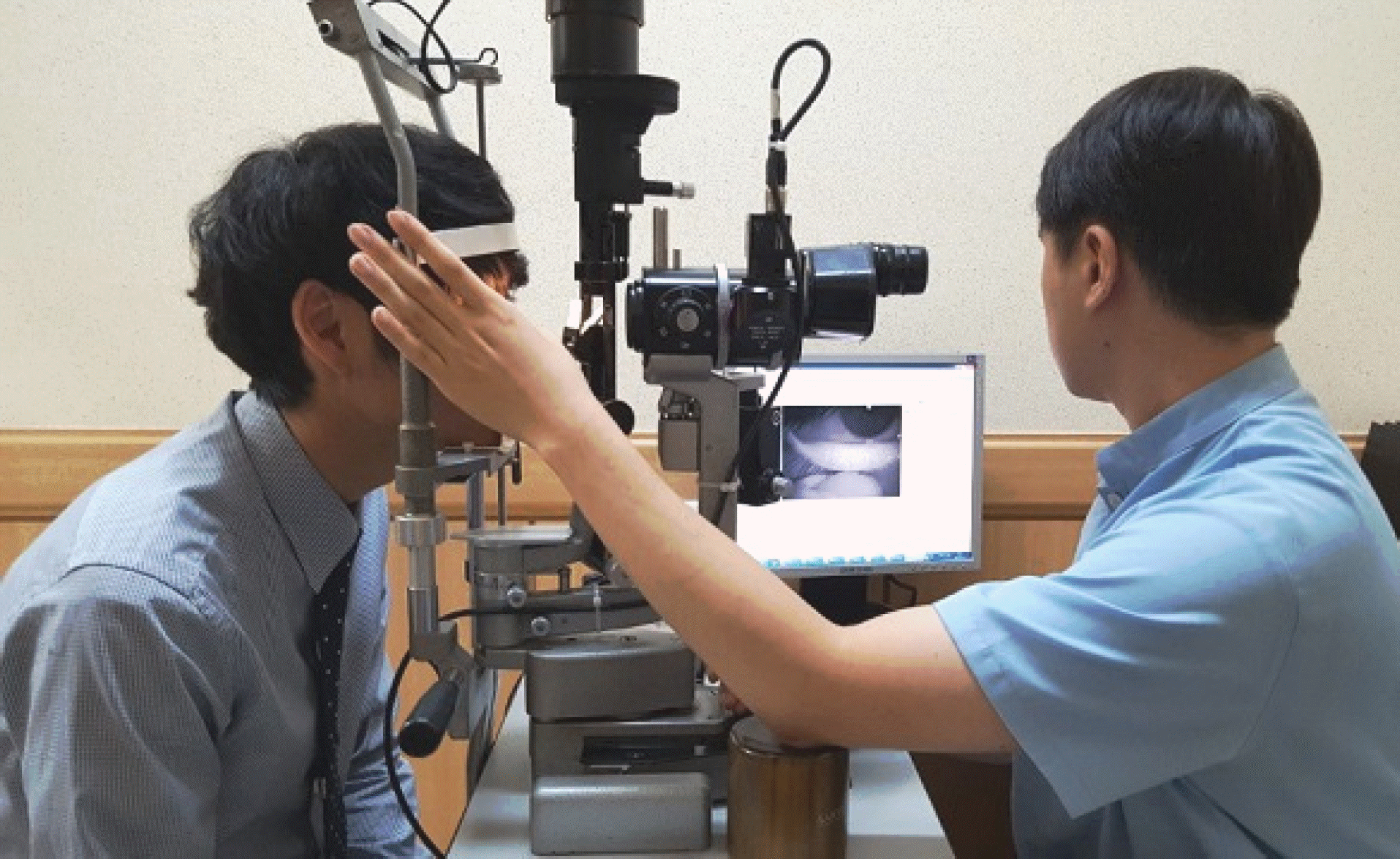 | Figure 1.Picture of performing indirect infrared meibography. Infrared-only charge-coupled device camera was fitted to a slit lamp. |
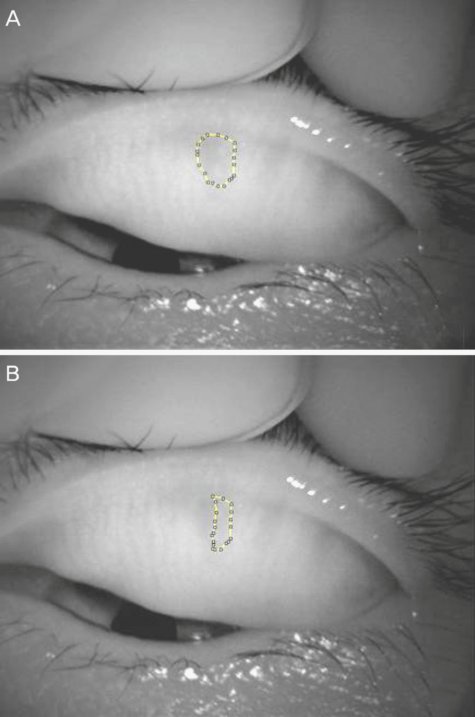 | Figure 2.Meibographies of one patient before incision and curettage of chalazion. (A) Dotted line represent dark area, (B) dotted line represent intact meibomian gland inside dark area. |
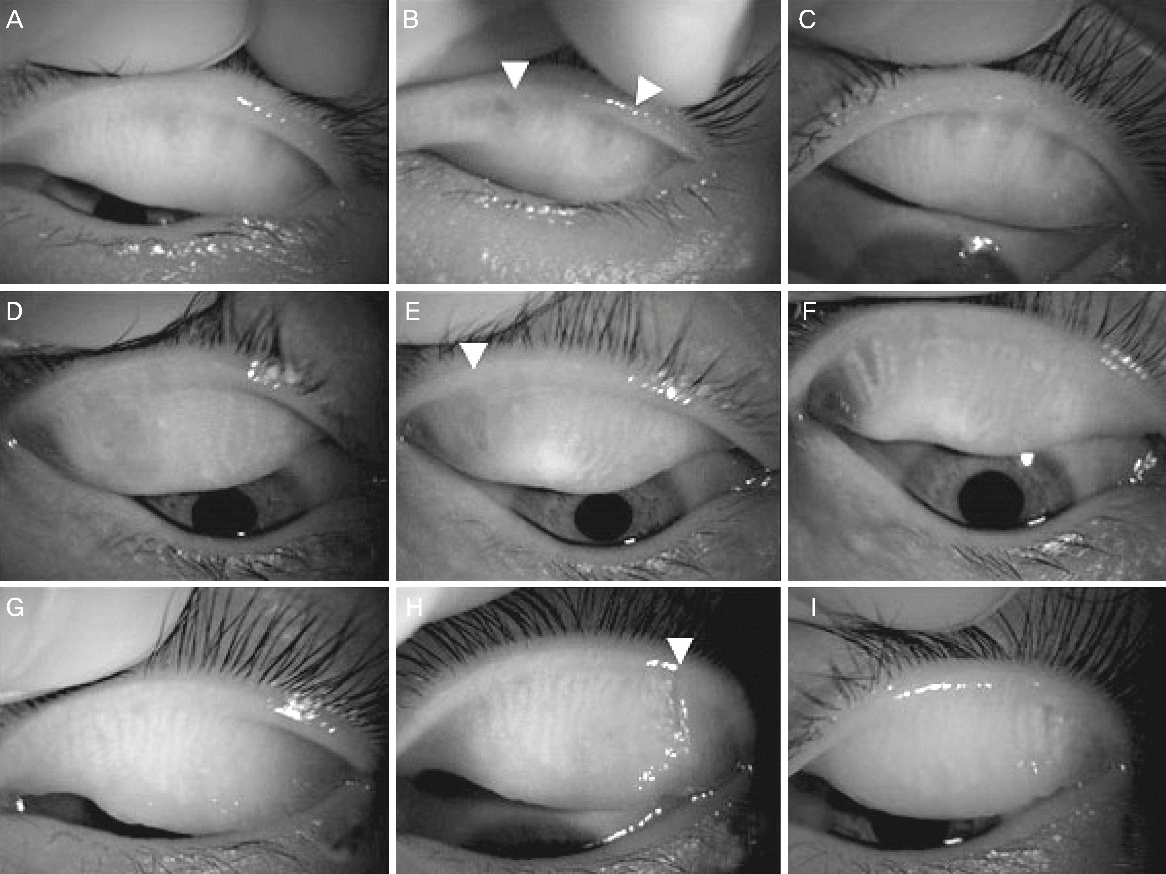 | Figure 3.Pictures of meibomian gland of three patients by indirect meibography. Before procedure, inflammatory change and partial meibomian gland drop out was observed (A, D, G). 1 week after the procedure, incision wound (white arrowheads) was found at the operated area (B, E, H). 1 month after the procedure, scar was formed at original lesion, and surrounding normal meibomian glands were observed (C, F, I). |
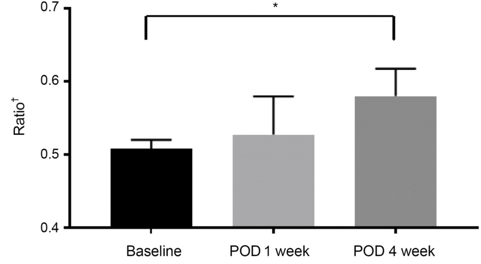 | Figure 4.Average ratio of ‘normal meibomian gland/normal meibomian gland and dark area’ with meibohrapgy by time after treatment. There was significant through 4 weeks (*p = 0.041, Paired t-test). POD = post operation date, time after incision & curettage. † Ratio of normal meibomian gland/dark area at meibography. |
Table 1.
Basic characteristics of patients with direction and location of chalazion lesion
| Patient | Age | Sex | Direction* | Location† |
|---|---|---|---|---|
| 1 | 57 | Male | Right | LL |
| 2 | 10 | Female | Left | UL |
| 3 | 6 | Male | Left | UL |
| 4 | 76 | Female | Right | LL |
| 5 | 35 | Female | Right | UL |
| 6 | 8 | Female | Right | UL |
| 7 | 64 | Female | Left | UL |
| 8 | 19 | Male | Right | UL |
| 9 | 42 | Male | Left | UL |
| 10 | 21 | Male | Right | UL |
| 11 | 30 | Male | Right | UL |
| 12 | 49 | Male | Right | UL |
| 13 | 8 | Male | Left | UL |
| 14 | 32 | Male | Left | UL |
| 15 | 52 | Female | Left | LL |
| 16 | 17 | Female | Right | UL |
Table 2.
Average of meiboscore, break up time, Schirmer test after incision & curettage of chalazion by time after treatment
| Baseline | POD 1 week | 4 week | p-value* | |
|---|---|---|---|---|
| Meiboscore | 1.36 ± 0.51 | 1.55 ± 0.69 | 1.27 ± 0.47 | 0.092 |
| BUT | 10.73 ± 2.45 | 11.36 ± 1.80 | 10.64 ± 2.33 | 0.068 |
| Schirmer† | 11.00 ± 4.69 | 10.91 ± 5.22 | 11.00 ± 4.58 | 0.972 |
 XML Download