Abstract
Purpose
To characterize the horizontal angular direction of lower eyelashes in different age groups of Korean patients.
Methods
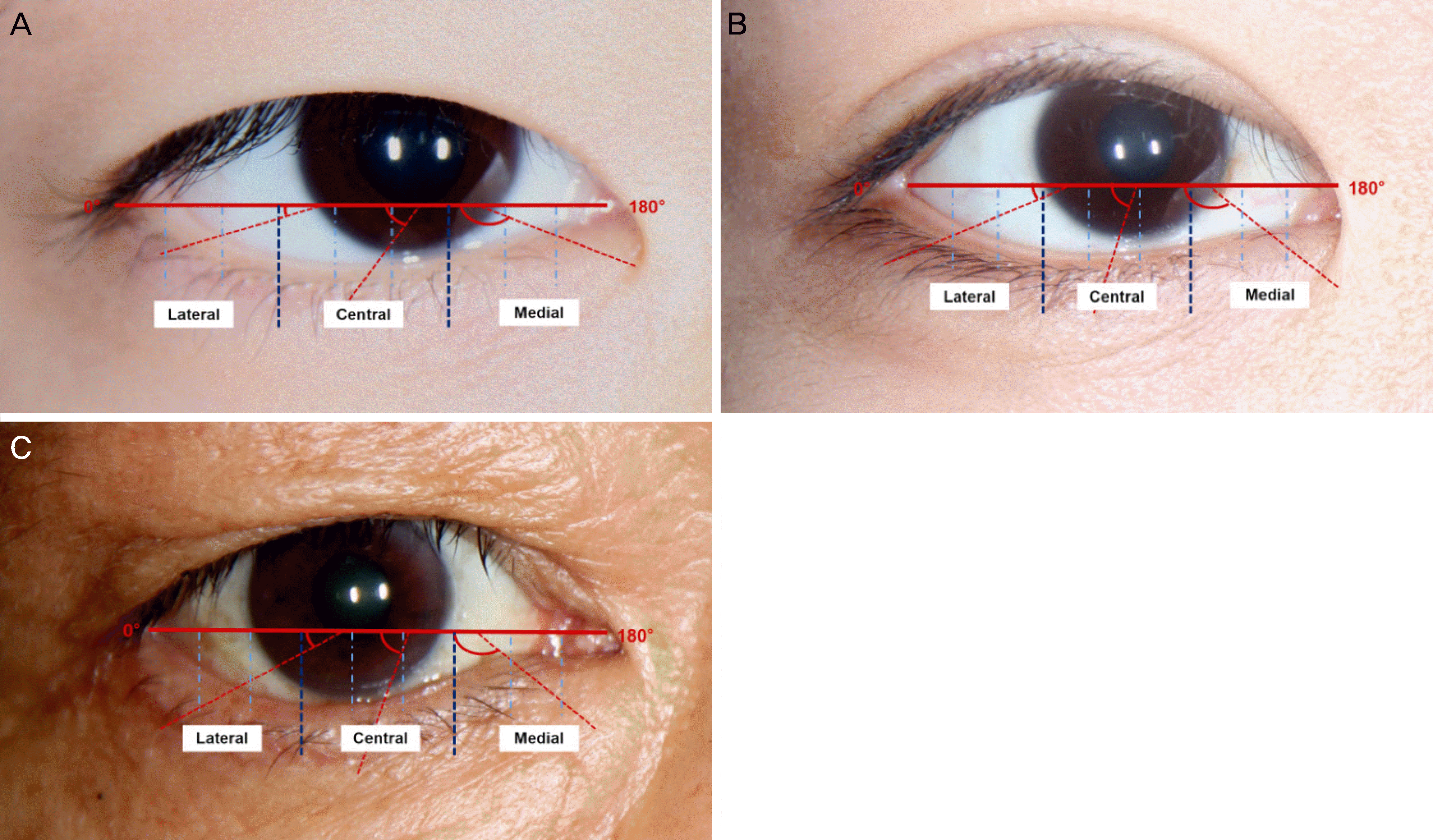
Digital photographs of 122 patients were obtained and the patients were divided into three groups involving children 6–19 years of age, adults 20–64 years of age, and older adults 65–85 years of age. Lower eyelashes were divided into medial, central, and lateral portions. Each portion was subdivided into three parts and the average value of the three parts was obtained for each portion. Horizontal angular directions tangential to the baseline between the medial and lateral canthus were measured and the data were compared between different portions and groups.
Results
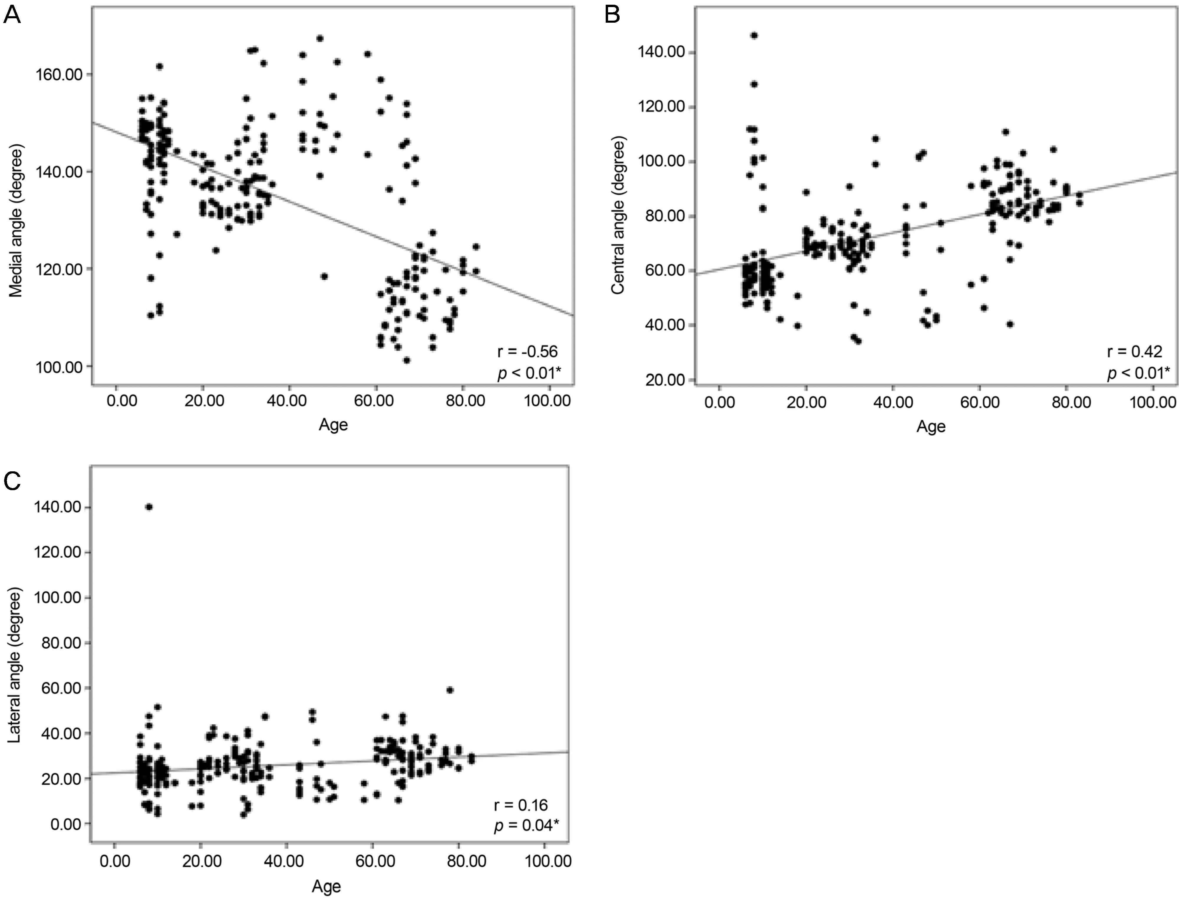
The mean horizontal angular directions of the lower eyelash in the medial portions were 142.4 ± 10.8° in children, 137.1 ± 13.8° in adults, and 120.4 ± 13.0° in older adults. There was a negative correlation with age (r = −0.56, p < 0.01). In central portions, the values were 62.8 ± 18.5°, 71.8 ± 14.5°, and 86.0 ± 10.5°, respectively; and in lateral portions they were 23.3 ± 13.7°,25.3 ± 9.3°, and 29.5 ± 8.1°. There were positive correlations with aging in the central and lateral portions (r = 0.42, p < 0.01; r = 0. 16, p = 0.04, respectively).
Conclusions
The mean horizontal angular direction of eye lashes decreases with age in the medial portion of the lower eyelid but increases in the central and lateral portions in Korean patients. This may be related to factors such as stretching of the eyelid and involutional horizontal eyelid laxity and orbicularis muscle changes.
Go to : 
References
1. Thibaut S, De Becker E, Caisey L, et al. Human eyelash characterization. Br J Dermatol. 2010; 162:304–10.

2. Na JI, Kwon OS, Kim BJ, et al. Ethnic characteristics of eyelashes: a comparative analysis in Asian and Caucasian females. British J Dermatol. 2006; 155:1170–6.

3. Hwang K. Surgical anatomy of the upper eyelid relating to upper blepharoplasty or blepharoptosis surgery. Anat Cell Biol. 2013; 46:93–100.

4. Langford JD, Linberg JV. A new physical finding in floppy eyelid syndrome. Ophthalmology. 1998; 105:165–9.

5. Malik KJ, Lee MS, Park DJ, Harrison AR. Lash ptosis in abdominal and acquired blepharoptosis. Arch Ophthalmol. 2007; 125:1613–5.
6. Lee TE, Lee JM, Lee H, et al. Lash ptosis and associated factors in Asians. Ann Plast Surg. 2010; 65:407–10.

7. Hahn S, Desai SC. Lower lid malposition: causes and correction. Facial Plast Surg Clin North Am. 2016; 24:163–71.
8. Murri M, Hamill EB, Hauck MJ, Marx DP. An update on lower lid blepharoplasty. Semin Plast Surg. 2017; 31:46–50.

9. Woo KI, Kim YD. Management of epiblepharon: state of the art. Curr Opin Ophthalmol. 2016; 27:433–8.
10. Procianoy F, Mendonça TB, Bins CA, Lang MP. Characterization of normal mediolateral angular direction of lower eyelid eyelashes in different age groups. Ophthal Plast Reconstr Surg. 2015; 31:332–3.

11. Glaser DA, Jones D, Carruthers J, et al. Epidemiologic analysis of change in eyelash characteristics with increasing age in a abdominal of healthy women. Dermatol Surg. 2014; 40:1208–13.
12. Kim JS, Jin SW, Hur MC, et al. The clinical characteristics and abdominal outcomes of epiblepharon in korean children: a 9-year experience. J Ophthalmol. 2014; 2014:156501.
13. Kim KH, Baek JS, Lee S, et al. Causes and surgical outcomes of lower eyelid retraction. Korean J Ophthalmol. 2017; 31:290–8.

14. Dunbar KE, Cox C, Heher KL, Kapadia MK. Lateral tarsal strip plus skin-muscle flap excision in the treatment of lower eyelid abdominal entropion. Orbit. 2017; 36:375–81.
15. Lee H, Park M, Chang M, et al. Clinical characteristics and abdominalness of the lateral tarsal strip and medial spindle procedure. Ann Plast Surg. 2015; 75:365–9.
16. Carter SR, Chang J, Aguilar GL, et al. Involutional entropion and ectropion of the Asian lower eyelid. Ophthal Plast Reconstr Surg. 2000; 16:45–9.

17. Marcet MM, Phelps PO, Lai JS. Involutional entropion: risk factors and surgical remedies. Curr Opin Ophthalmol. 2015; 26:416–21.
Go to : 
 | Figure 1.Measurement of angle of cilia in 3 different areas. (A) Child. (B) Adult. (C) Senile group. |
 | Figure 2.Correlation of age with angle of cilia in different portions.(A) Medial, (B) central, and (C) lateral portions. * Simple linear regression test. |
Table 1.
Demographics and measurements of angle of cilia
| Total (n = 244 eyes) | Child (n = 78 eyes) | Adult (n = 110 eyes) | Senile (n = 56 eyes) | p-value* | |
|---|---|---|---|---|---|
| Age (years) | 35.8 ± 24.7 (6–83) | 9.1 ± 2.6 (6–18) | 36.7 ± 13.8 (20–64) | 71.1 ± 5.2 (65–83) | <0.01 |
| Angle of cilia | |||||
| Medial | 135.2 ± 14.4 | 142.4 ± 10.8 | 137.1 ± 13.8 | 120.4 ± 13.0 | <0.01 |
| (101.2–167.4) | (110.4–161.6) | (104.3–167.4) | (101.2–155.1) | ||
| Central | 72.4 ± 18.1 | 62.8 ± 18.5 | 71.8 ± 14.5 | 86.0 ± 10.5 | <0.01 |
| (34.1–146.3) | (39.7–146.3) | (34.1–108.4) | (40.3–110.9) | ||
| Lateral | 25.4 ± 10.7 | 23.3 ± 13.7 | 25.3 ± 9.3 | 29.5 ± 8.1 | 0.04 |
| (3.9–140.3) | (4.3–140.3) | (3.9–49.4) | (10.4–59.1) |


 PDF
PDF ePub
ePub Citation
 Print
Print


 XML Download