Abstract
Methods
The medical records of patients who were diagnosed with metastatic orbital tumors at Asan Medical Center, Seoul, Republic of Korea, from May 2010 to May 2017, were retrospectively reviewed.
Results
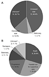
A total of 27 patients were studied, including 14 (51.9%) males. The mean age at diagnosis was 54.1 years and the mean follow-up period was 12.7 months. The most common primary tumor site was the breast (six patients, 46.2%) in females and the liver (five patients, 35.7%) in males. Proptosis (12 patients, 44.4%) was the most common complaint, with the other complaints including periorbital swelling, palpable masses and diplopia. Orbital imaging studies showed that the most common sites of orbital involvement were the orbital wall (nine patients, 33.3%) and the extraocular muscle (nine patients, 33.3%). Pathological diagnoses were performed in 10 patients (37.0%), 3 of these whom underwent a fine needle aspiration biopsy for intraconal lesions. Local and systemic therapies were conducted using a multidisciplinary approach, but the size of orbital tumors increased in 12 (44.4%) of 27 patients. Eight (29.6%) patients died because of systemic metastasis, seven (25.9%) patients were referred to other institutions for palliative treatment, and five (18.5%) patients showed malignant progression despite treatment.
Conclusions
Based on the varying clinical features of metastatic orbital tumors, it is important to carefully diagnose patients, especially those with a history of cancer. Breast cancer is the most common primary cancer, and liver cancer is also common because of the high prevalence of hepatocellular carcinoma in the Republic of Korea. Clinical diagnosis may be possible using patient history and/or imaging study data, but pathological confirmation may be necessary in some cases. The treatment options can be determined by the type of primary cancer and the general condition, but the overall prognosis is poor.
Figures and Tables
Figure 1
Orbital metastasis from breast cancer. (A, B) Magnetic resonance images revealed thickened of left inferior rectus muscle and infiltrative lesion extending to left inferior oblique muscle and overlying lower eyelid. (C) Clinical photos showed supraduction limitation in the left eye. (D) HESS screen test also showed supraduction limitation in left eye and left hypotropia. (E) Pathologic study of left inferior rectus muscle biopsy demonstrated infiltration of metastatic cells with scant cytoplasm and hyperchromatic nuclei (H&E stain, original magnification ×200).

Figure 2
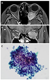
Magnetic resonance imaging of the metastatic orbital tumor from thymic origin neuroendocrine carcinoma. (A) coronal view (B) axial view, which showed an enhancing mass with lobulating contour (2.6 × 1.9 cm) in the left retrobulbar area. (C) Smear from fine-needle aspiration showed numerous spindle cells with hyperchromatic nuclei (H&E stain, original magnification ×400).

Figure 3
Clinical course of total patients. (A) Orbital lesion and (B) general conditions after treatment for metastatic orbital tumors (*Loss to follow-up).

References
1. Finger PT. Radiation therapy for orbital tumors: concepts, current use, and ophthalmic radiation side effects. Surv Ophthalmol. 2009; 54:545–568.

2. Bloch RS, Gartner S. The incidence of ocular metastatic carcinoma. Arch Ophthalmol. 1971; 85:673–675.

3. Albert DM, Rubenstein RA, Scheie HG. Tumor metastasis to the eye. I. Incidence in 213 adult patients with generalized malignancy. Am J Ophthalmol. 1967; 63:723–726.
4. Valenzuela AA, Archibald CW, Fleming B, et al. Orbital metastasis: clinical features, management and outcome. Orbit. 2009; 28:153–159.

5. Civit T, Colnat-Coulbois S, Freppel S. Orbital metastasis. Neurochirurgie. 2010; 56:148–151.
7. Shields JA, Shields CL, Scartozzi R. Survey of 1264 patients with orbital tumors and simulating lesions: The 2002 Montgomery Lecture, part 1. Ophthalmology. 2004; 111:997–1008.
8. Ahmad SM, Esmaeli B. Metastatic tumors of the orbit and ocular adnexa. Curr Opin Ophthalmol. 2007; 18:405–413.

9. Shikishima K, Kawai K, Kitahara K. Pathological evaluation of orbital tumours in Japan: analysis of a large case series and 1379 cases reported in the Japanese literature. Clin Exp Ophthalmol. 2006; 34:239–244.

10. Tomizawa Y, Ocque R, Ohori NP. Orbital metastasis as the initial presentation of invasive lobular carcinoma of breast. Intern Med. 2012; 51:1635–1638.

11. Gupta S, Bhatt VR, Varma S. Unilateral orbital pain and eyelid swelling in a 46-year-old woman: orbital metastasis of occult invasive lobular carcinoma of breast masquerading orbital pseudotumour. BMJ Case Rep. 2011; 2011:pii: bcr1220103580.

12. Abbruzzese JL, Abbruzzese MC, Lenzi R, et al. Analysis of a diagnostic strategy for patients with suspected tumors of unknown origin. J Clin Oncol. 1995; 13:2094–2103.

13. Papathanassiou M, Nikita E, Theodossiadis P, Vergados I. Orbital metastasis secondary to breast cancer mimicking thyroid-associated ophthalmopathy. Clin Exp Optom. 2010; 93:368–369.

14. Kong BD, Lee TW. Clinical analysis of metastatic intraocular malignancy. J Korean Ophthalmol Soc. 1999; 40:2928–2934.
15. Shields CL, Shields JA, Peggs M. Tumors metastatic to the orbit. Ophthal Plast Reconstr Surg. 1988; 4:73–80.

16. Goldberg RA, Rootman J, Cline RA. Tumors metastatic to the orbit: a changing picture. Surv Ophthalmol. 1990; 35:1–24.

17. Tijl J, Koornneef L, Eijpe A, et al. Metastatic tumors to the orbit--management and prognosis. Graefes Arch Clin Exp Ophthalmol. 1992; 230:527–530.

18. Shields JA, Shields CL, Brotman HK, et al. Cancer metastatic to the orbit: the 2000 Robert M. Curts Lecture. Ophthal Plast Reconstr Surg. 2001; 17:346–354.
19. Yan J, Gao S. Metastatic orbital tumors in southern China during an 18-year period. Graefes Arch Clin Exp Ophthalmol. 2011; 249:1387–1393.

20. Günalp I, Gündüz K. Metastatic orbital tumors. Jpn J Ophthalmol. 1995; 39:65–70.
21. Font RL, Ferry AP. Carcinoma metastatic to the eye and orbit III. A clinicopathologic study of 28 cases metastatic to the orbit. Cancer. 1976; 38:1326–1335.
22. Öberg K, Hellman P, Ferolla P, et al. Neuroendocrine bronchial and thymic tumors: ESMO Clinical Practice Guidelines for diagnosis, treatment and follow-up. Ann Oncol. 2012; 23:Suppl 7. vii120–vii123.

23. Goldberg RA, Rootman J. Clinical characteristics of metastatic orbital tumors. Ophthalmology. 1990; 97:620–624.

24. Green S, Som PM, Lavagnini PG. Bilateral orbital metastases from prostate carcinoma: case presentation and CT findings. AJNR Am J Neuroradiol. 1995; 16:417–419.
25. Sa HS, Oh DE, Kim YD. Fine needle aspiration biopsy of posterior orbital tumors. J Korean Ophthalmol Soc. 2005; 46:1435–1440.
26. Kennerdell JS, Dekker A, Johnson BL, Dubois PJ. Fine-needle aspiration biopsy. Its use in orbital tumors. Arch Ophthalmol. 1979; 97:1315–1317.


 PDF
PDF ePub
ePub Citation
 Print
Print


 XML Download