

 PDF
PDF ePub
ePub Citation
 Print
Print


Abstract
Purpose
To report the first case of steroid sulfatase (STS) gene deletion, confirmed by multiplex ligation-dependent probe amplification (MLPA) analysis in identical twins with pre-Descemet corneal dystrophy associated with X-linked ichthyosis.
Case summary
19-year old identical twin brothers with itching senses and hereditary thick scaly skin of the extremity and trunk visited our dermatologic clinic. Upon visiting, an ophthalmologic consultation with anterior segment examination showed diffuse punctate corneal opacities in the pre-Descemet layer. On MLPA analysis of the identical twin brothers, a definitive diagnosis of X-linked ichthyosis was made by identifying STS gene deletion.
Figures and Tables
Figure 1
Histopathological picture of patient's skin. The black arrowheads indicate hyperkeratosis (Hematoxylin-eosin stain; original magnification: ×100).
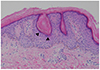
Figure 2
Slit-lamp microscopic photograph of affected patient. (A) Slit-lamp photograph of the cornea in diffuse illumination show no definite corneal lesion. (B) Slit-lamp photograph in slit illumination show diffuse small punctate opacities (red dot line circle) observed in stroma and anterior to Descemet's membrane.

Figure 3
All of specular microscopy show normal endothelial cell density and morphology. (A) Twin older brother. (B) Twin younger brother. CD =cell density; SD = standard deviation; CV = coefficient of variation; 6A = percentage of hexagonal cell; AVE = average cell area; MAX = maximum cell area; MIN = minimum cell area; R = right; L = left.
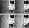
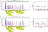
Figure 4
The Multiplex ligation-dependent probe amplification (MLPA) analysis of patient. (A) Twin older brother, and (B) Twin younger brother. Left graph (blue graph-patient, red graph-normal control) and right scatter plot (in dot line box) of each case show deletion of STS from exon 1 to exon 10 except exon 3 and contiguous HDHD1A upstream deletion. STS, steroid sulfatase.
References
1. Wells RS, Kerr CB. Genetic classification of ichthyosis. Arch Dermatol. 1965; 92:1–6.
2. Wells RS, Kerr CB. Clinical features of autosomal dominant and sex-linked ichthyosis in an English population. Br Med J. 1966; 1:947–950.
3. Bale SJ, Doyle SZ. The genetics of ichthyosis: a primer for epidemiologists. J Invest Dermatol. 1994; 102:49S–50S.
4. Casaroli Marano RP, Ortiz Stradtmann MA, Uxo M, Iglesias E. Ocular findings associated with congenital X-linked ichthyosis. Ann Ophthalmol. 1991; 23:167–172.
5. Haritoglou C, Ugele B, Kenyon KR, Kampik A. Corneal manifestations of X-linked ichthyosis in two brothers. Cornea. 2000; 19:861–863.
6. Kempster RC, Hirst LW, de la Cruz Z, Green WR. Clinicopathologic study of the cornea in X-linked ichthyosis. Arch Ophthalmol. 1997; 115:409–415.
7. Hernández-Martín A, González-Sarmiento R, De Unamuno P. X-linked ichthyosis: an update. Br J Dermatol. 1999; 141:617–627.
8. Reed MJ, Purohit A, Woo LW, et al. Steroid sulfatase: molecular biology, regulation, and inhibition. Endocr Rev. 2005; 26:171–202.
9. Hung C, Ayabe RI, Wang C, et al. Pre-descemet corneal dystrophy and X-linked ichthyosis associated with deletion of Xp22.31 containing the STS gene. Cornea. 2013; 32:1283–1287.
10. Jeong CK, Hong JS, Lee TH, Lee HY. A case of corneal opacity in X-linked ichtyosis patient. J Korean Ophthalmol Soc. 1996; 37:1085–1089.
11. Lykkesfeldt G, Hoyer H, Jbsen HH, Bandrup F. Steroid sulfatase deficiency disease. Clin Genet. 1985; 28:231–237.
12. Sever RJ, Frost P, Weinstein G. Eye changes in ichthyosis. JAMA. 1968; 206:2283–2286.
13. Macsai MS, Doshi H. Clinical pathologic correlation of superficial corneal opacities in X-linked ichthyosis. Am J ophthalmol. 1994; 118:477–484.
14. Costagliola C, Fabbrocini G, Illiano GM, et al. Ocular findings in X-linked ichthyosis: a survey on 38 cases. Ophthalmologica. 1991; 202:152–155.
15. Fernandes NF, Janniger CK, Schwartz RA. X-linked ichthyosis: an oculocutaneous genodermatosis. J Am Acad Dermatol. 2010; 62:480–485.
16. Schwartz M, Dunø M. Improved molecular diagnosis of dystrophin gene mutations using the multiplex ligation-dependent probe amplification method. Genet Test. 2004; 8:361–367.
17. Kim GH, Lee BH, Yoo HW. MLPA application in genetic testing. J Genet Med. 2009; 6:146–154.
 XML Download