

 PDF
PDF ePub
ePub Citation
 Print
Print


Abstract
Purpose
To determine the association between optic disc tilt and torsion of glaucomatous and fellow eyes of unilateral normal-tension glaucoma (NTG) patients and normal.
Methods
We measured optic disc tilt and torsion of 23 unilateral NTG patients and 23 normal controls by analyzing fundus photography and compared 3 groups. We also measured retinal nerve fiber layer (RNFL) thickness through optical coherence tomography in 23 unilateral NTG patients and compared the findings with those of normal eyes.
Results
The mean values of optic disc tilt ratio and torsion degree in glaucomatous eyes were 1.17 ± 0.19° and 15.57 ± 8.16°, respectively, while those in fellow eyes were 1.10 ± 0.10° and 8.26 ± 5.20°. There was no significant difference in tilt ratio (p = 0.109), but there was a significant difference in torsion degree (p = 0.001). The mean values of optic disc tilt ratio and optic disc torsion in the controls were 1.11 ± 0.07° and 3.25 ± 2.69°, respectively. Also, there was no significant difference in optic disc tilt ratio (p = 0.601), but a significant difference in optic disc torsion between fellow eyes and controls (p < 0.001). The RNFL thickness of the same torsion direction in unilateral NTG eyes was measured to be 49.35 ± 17.18 µm smaller than the normal value (mean RNFL thickness: 71.91 ± 16.92 µm). Reduced RNFL thickness of the same torsion direction between glaucomatous eyes and fellow eyes was significantly different (p < 0.001). In addition, it was confirmed that RNFL thickness was significantly decreased according to the degree of disc torsion (p = 0.024).
Conclusions
The optic disc torsion showed a significant difference between glaucomatous and fellow eyes in unilateral NTG patients and normal controls. Also, the RNFL thickness significantly decreased according to the degree of the optic disc torsion. Therefore, fellow eyes of unilateral NTG patients need to be carefully monitored for the progression of glaucoma.
Figures and Tables
Figure 1
Definitions of optic disc tilt and torsion degree with Image J software. Optic disc tilt ratio was defined as the ratio between the longest diameter (LD) and shortest diameter (SD) of the optic disc. Optic disc torsion degree was measured between the LD and vertical meridian (VM) of the optic disc (θ).
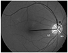
Figure 2
The bar chart of the optic disc tilt ratio and torsion degree. Comparisons of the optic disc tilt ratio (A) and torsion degree (B) between glaucomatous eyes and fellow eyes of 23 patients with unilateral normal-tension glaucoma (NTG) patients, and 46 normal controls. G-NTG = glaucomatous eyes of unilateral normal-tension glaucoma; Fellow eye-NTG = contralateral normal eyes of unilateral normal-tension glaucoma.
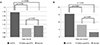
Figure 3
Scatter map between optic disc torsion and retinal nerve fiber layer (RNFL) thickness of the same direction. Scatterplots showing the relationship between the optic disc torsion degree and the same directional RNFL thickness (A) and the difference between the same directional RNFL thickness and normal values (B). Pearson correlation coefficient R values are shown. S_RNFL = same directional RNFL thickness (µm); S_RNFL_D = difference between the same directional RNFL thickness and normal values (µm).
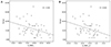
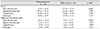
Table 1
The results of eye examinations and demographics in 23 patients with unilateral normal-tension glaucoma (NTG) and 46 eyes of 23 normal controls
Table 2
The optic disc tilt ratio and torsion degree of 23 patients with unilateral normal-tension glaucoma (NTG) and 46 eyes of 23 normal controls

References
1. Shields MB. Normal-tension glaucoma: is it different from primary open-angle glaucoma? Curr Opin Ophthalmol. 2008; 19:85–88.
2. Yamazaki Y, Drance SM. The relationship between progression of visual field defects and retrobulbar circulation in patients with glaucoma. Am J Ophthalmol. 1997; 124:287–295.
3. Park HY, Lee K, Park CK. Optic disc torsion direction predicts the location of glaucomatous damage in normal-tension glaucoma patients with myopia. Ophthalmology. 2012; 119:1844–1851.
4. Park HY, Lee KI, Lee K, et al. Torsion of the optic nerve head is a prominent feature of normal-tension glaucoma. Invest Ophthalmol Vis Sci. 2014; 56:156–163.
5. Sohn SW, Kee CW. Influence of myopia on the progression of normal tension glaucoma. J Korean Ophthalmol Soc. 2007; 48:527–534.
6. Mitchell P, Hourihan F, Sandbach J, Wang JJ. The relationship between glaucoma and myopia: the Blue Mountains Eye Study. Ophthalmology. 1999; 106:2010–2015.
7. McBrien NA, Gentle A. Role of the sclera in the development and pathological complications of myopia. Prog Retin Eye Res. 2003; 22:307–338.
8. Yang YS, Koh JW. Choroidal blood flow change in eyes with high myopia. Korean J Ophthalmol. 2015; 29:309–314.
9. Lee KS, Lee JR, Kook MS. Optic disc torsion presenting as unilateral glaucomatous-appearing visual field defect in young myopic Korean eyes. Ophthalmology. 2014; 121:1013–1019.
10. Cho HK, Suh W, Kee C. Visual and structural prognosis of the untreated fellow eyes of unilateral normal tension glaucoma patients. Graefes Arch Clin Exp Ophthalmol. 2015; 253:1547–1555.
11. Rudnicka AR, Mt-lsa S, Owen CG, et al. Variation in primary open-angle glaucoma prevalence by age, gender, and race: a Bayesian meta-analysis. Invest Ophthalmol Vis Sci. 2006; 47:4254–4261.
12. Stein JD, Kim DS, Niziol LM, et al. Differences in rates of glaucoma among Asian Americans and other racial groups, and among various Asian ethnic groups. Ophthalmology. 2011; 118:1031–1037.
13. Kim CS, Seong GJ, Lee NH, et al. Prevalence of primary open-angle glaucoma in central South Korea the Namil study. Ophthalmology. 2011; 118:1024–1030.
14. Kim JM, Jeoung JW, Bitrian E, et al. Comparison of clinical characteristics between Korean and Western normal-tension glaucoma patients. Am J Ophthalmol. 2013; 155:852–857.
15. Kato T, Meguro A, Nomura E, et al. Association study of genetic variants on chromosome 7q31 with susceptibility to normal tension glaucoma in a Japanese population. Eye (Lond). 2013; 27:979–983.
16. Takamoto M, Kaburaki T, Mabuchi A, et al. Common variants on chromosome 9p21 are associated with normal tension glaucoma. PLoS One. 2012; 7:e40107.
17. Shi D, Funayama T, Mashima Y, et al. Association of HK2 and NCK2 with normal tension glaucoma in the Japanese population. PLoS One. 2013; 8:e54115.
18. Kim DM, Hwang US, Park KH, Kim SH. Retinal nerve fiber layer thickness in the fellow eyes of normal-tension glaucoma patients with unilateral visual field defect. Am J Ophthalmol. 2005; 140:165–166.
 XML Download