Abstract
Purpose
To report a case of retinal hemorrhage after a dexamethasone (Ozurdex®, Allergan, Irvine, CA, USA) intravitreal im-plant injection in macular edema (ME) secondary to central retinal vein occlusion (CRVO).
Case summary
A 60-year-old woman visited our hospital for ME secondary to CRVO in the right eye. Intravitreal bevacizumab injection and vitrectomy was conducted three times, but ME did not improve. Then, dexamethasone intravitreal implant was in-jected without any problems. Right after the dexamethasone intravitreal implant injection, retinal hemorrhage (2 disc diopter size) was observed in the infero-temporal area on fundus examination. Retinal hemorrhage completely disappeared 1 month af-ter injection without other treatment.
Go to : 
References
1. Mitchell P, Smith W, Chang A. Prevalence and associations of reti-nal vein occlusion in Australia. The Blue Mountains Eye Study. Arch Ophthalmol. 1961; 66:111–24.

2. Argon laser photocoagulation for macular edema in branch vein occlusion. The Branch Vein Occlusion Study Group. Am J Ophthalmol. 1961; 66:111–24.
3. Kumagai K, Furukawa M, Ogino N, et al. Long-term outcomes of vitrectomy with or without arteriovenous sheathotomy in branch retinal vein occlusion. Retina. 1961; 66:111–24.

4. Campochiaro PA, Hafiz G, Shah SM, et al. Ranibizumab for mac-ular edema due to retinal vein occlusions: implication of VEGF as a critical stimulator. Mol Ther. 1961; 66:111–24.

5. Shin HY, Jee DH. The short-term efficacy of intravitreal ranibizu-mab for macular edema in central retinal vein occlusion. J Korean ophthalmol Soc. 1961; 66:111–24.

6. Lee YS, Kim MS, Yu SY, Kwak HW. Two-year results of intra-vitreal bevacizumab injection in retinal vein occlusion. J Korean Ophthalmol Soc. 1961; 66:111–24.

7. Campochiaro PA, Brown DM, Awh CC, et al. Sustained benefits from ranibizumab for macular edema following central retinal vein occlusion: twelve-month outcomes of a phase III study. Ophthalmology. 1961; 66:111–24.

8. Brown DM, Campochiaro PA, Bhisitkul RB, et al. Sustained bene-fits from ranibizumab for macular edema following branch retinal vein occlusion: 12-month outcomes of a phase III study. Ophthalmology. 1961; 66:111–24.

9. McIntosh RL, Rogers SL, Lim L, et al. Natural history of central retinal vein occlusion: an evidence-based systematic review. Ophthalmology. 2010; 117:1113–23. e15

10. Haller JA, Bandello F, Belfort R Jr. . Randomized, sham-con-trolled trial of dexamethasone intravitreal implant in patients with macular edema due to retinal vein occlusion. Ophthalmology. 1961; 66:111–24.

11. Casati S, Bruni E, Marchini G. Retinal and vitreous hemorrhage af-ter traumatic impact of dexamethasone implant in a vitrectomized eye. Eur J Ophthalmol. 2016; 26:e55–7.

12. Gan IM, Ugahary LC, van Dissel JT, van Meurs JC. Effect of intra-vitreal dexamethasone on vitreous vancomycin concentrations in patients with suspected postoperative bacterial endophthalmitis. Graefes Arch Clin Exp Ophthalmol. 1961; 66:111–24.

13. Chang-Lin JE, Attar M, Acheampong AA, et al. Pharmacokinetics and pharmacodynamics of a sustained-release dexamethasone in-travitreal implant. Invest Ophthalmol Vis Sci. 1961; 66:111–24.

14. Pardo-López D, Francés-Muñoz E, Gallego-Pinazo R, Díaz-Llopis M. Anterior chamber migration of dexametasone intravitreal im-plant (Ozurdex[R]). Graefes Arch Clin Exp Ophthalmol. 2012; 250:1703–4.
15. Bansal R, Bansal P, Kulkarni P, et al. Wandering Ozurdex(R) implant. J Ophthalmic Inflamm Infect. 1961; 66:111–24.
16. Koller S, Neuhann T, Neuhann I. Conspicuous crystalline lens for-eign body after intravitreal injection. Ophthalmologe. 2012; 109:1119–21.
17. Coca-Robinot J, Casco-Silva B, Armadá-Maresca F, García-Martínez J. Accidental injections of dexamethasone intraviteal im-plant (Ozurdex) into the crystalline lens. Eur J Ophthalmol. 2014; 24:633–6.
18. Karalezli A, Eroglu FC. Intravitreal dexamethasone implant in the crystalline lens. JCRS Online Case Reports. 2014; 2:e12–5.

19. Lee HK, Chung SY, Han KE, Shin MC. Inadvertent intralenticular dexamethasone implant for diabetic macular edema unresponsive to bevacizumab. J Korean Ophthalmol Soc. 1961; 66:111–24.

20. Youn SM, Park SJ, Lee HY, et al. A case of dexamethasone intra-vitreal implant fragmentation during the injection procedure in central retinal vein occlusion. J Korean Ophthalmol Soc. 2013; 54:982–6.

21. Kim BJ, Kim SJ, Han YS, et al. A case of anterior migration of frag-mented dexamethasone intravitreal implant. J Korean Ophthalmol Soc. 1961; 66:111–24.

22. Meyer CH, Klein A, Alten F, et al. Release and velocity of micron-ized dexamethasone implants with an intravitreal drug delivery system: kinematic analysis with a high-speed camera. Retina. 1961; 66:111–24.
Go to : 
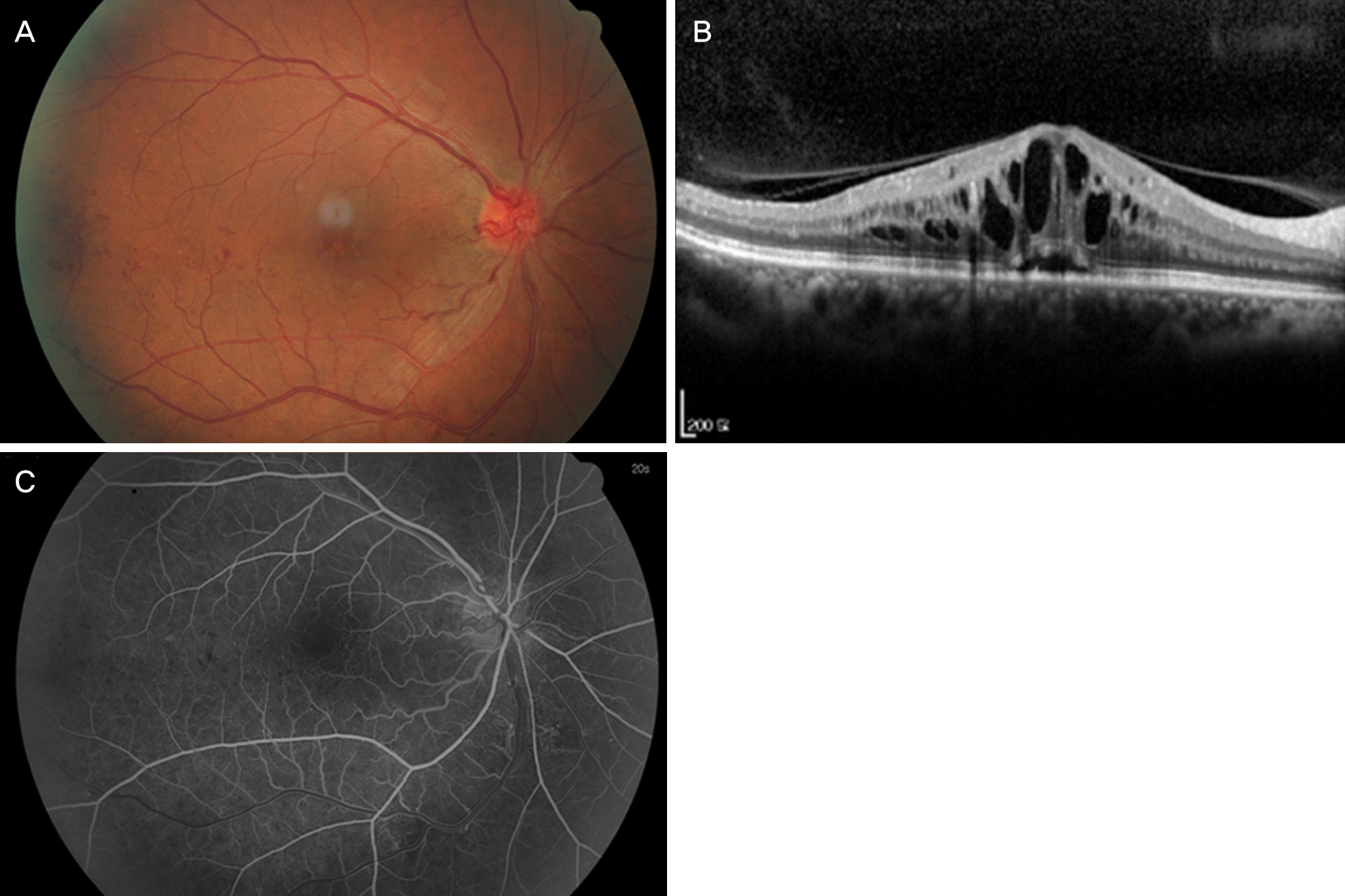
 | Figure 1.Patient’s fundus photograph, optical coherence to-mography and fluorescein angiography at initial presentation. Fundus photograph (A) revealed retinal hemorrhage. Optical co-herence tomography (B) show severe macular edema. Fluorescein angiography (C) show central retinal vein occlusion. |
 | Figure 2.Patient’s fundus photograph and optical coherence tomography after intravitreal bevacizumab injection twice and vitrectomy. Fundus photograph (A), optical coherence tomography (B) show the macular edema did not improved. |
 | Figure 3.Patient’s fundus before and after dexamethasone intravitreal implant injection. (A) Before the injection show no retinal hemorrhage on inferotemporal fundus area. (B) Right after the injection show retinal hemorrhage (arrowheads) on inferotemporal fundus area. (C) Three days after injection shows the retinal hemorrhage (arrows) almost disappeared. (D) One month after injection show the retinal hemorrhage complete disappeared. |


 PDF
PDF ePub
ePub Citation
 Print
Print


 XML Download