

 PDF
PDF ePub
ePub Citation
 Print
Print


Abstract
Purpose
To evaluate the histopathological changes of anterior capsule and lens epithelial cells in various types of cataract.
Methods
Patients scheduled for cataract surgery of phacoemulsification with intraocular lens implantation were enrolled in this study. Anterior capsule tissues sized 5 mm were obtained at the time of continuous curvilinear capsulorhexis during surgery. Histological examination of the obtained tissue was performed by transmission electron microscope.
Results
Nuclear cataract showed a uniform cuboidal monolayer of epithelial cells firmly attached to the anterior capsule. But, the mitochondria, Golgi apparatus, and endoplasmic reticulum were damaged and replaced with vacuoles. Anterior subcapsular cataract showed multilayers of epithelial cells with irregular intracellular structures. Epithelial cells of mature cataract were severely damaged and detached from the anterior capsule, accompanied by expansion of intra-cellular space and a large amount of vacuoles. Epithelial cells were irregular and severely damaged, and intracellular structures were hardly observed in traumatic cataract. Deposition of pseudoexfoliation materials on the anterior capsule was observed in pseudoexfoliation cataract.
Figures and Tables
Figure 1
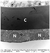
Transmission electron microscopy findings of normal lens. Uniform cuboidal monolayer epithelial cells are firmly attached to the anterior capsule (C). Regularly round shaped nucleus (N) with mitochondria, Golgi apparatus, and endoplasmic reticulum is observed in the cytoplasm (arrows).
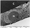
Figure 2
Transmission electron microscopy findings of nuclear cataract. Uniform cuboidal monolayer epithelial cells are firmly attached to the anterior capsule (C). Although nucleus (N) showing regular round shape, mitochondria, Golgi apparatus, and endoplasmic reticulum are damaged and replaced with vacuoles (arrows).
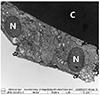
Figure 3
Transmission electron microscopy findings of anterior subcapsular cataract. Epithelial cells are elongated and are showing multilayer form interrupted by lens fibers. Nucleus (N) shows irregularity. Mitochondria, Golgi apparatus, and endoplasmic reticulum are damaged and hard to observe. C = anterior capsule.
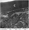
Figure 4
Transmission electron microscopy findings of mature cataract. Epithelial cells are severely damage and detached from the anterior capsule (C) followed by expansion of intra-cellular space and large amount of vacuoles (V) accumulation. Nucleus, Mitochondria, Golgi apparatus, and endoplasmic reticulum are damaged and hard to observe.
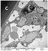
Figure 5
Transmission electron microscopy findings of traumatic cataract. Epithelial cells are showing its irregularity and severely damaged and also hard to observe. Nucleus, Mitochondria, Golgi apparatus, and endoplasmic reticulum are also damaged and hard to observe. C = anterior capsule.
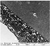
Figure 6
Transmission electron microscopy findings of pseudoexfoliation cataract. Uniform cuboidal monolayer epithelial cells are firmly attached to the anterior capsule (C). Regularly round shaped nucleus (N) with mitochondria, Golgi apparatus, and endoplasmic reticulum are observed in the cytoplasm (arrow). Pseudoexfoliation material deposition is observed on the anterior capsule (*).
References
1. Danysh BP, Duncan MK. The lens capsule. Exp Eye Res. 2009; 88:151–164.
2. Fisher RF. The structure and function of basement membrane (lens capsule) in relation to diabetes and cataract. Trans Ophthalmol Soc U K. 1985; 104(Pt 7):755–759.
3. Bhat SP. The ocular lens epithelium. Biosci Rep. 2001; 21:537–563.
4. Mathias RT, Rae JL, Baldo GJ. Physiological properties of the normal lens. Physiol Rev. 1997; 77:21–50.
5. Fischbarg J, Diecke FP, Kuang K, et al. Transport of fluid by lens epithelium. Am J Physiol. 1999; 276(3 Pt 1):C548–C557.
6. Giblin FJ, Chakrapani B, Reddy VN. Glutathione and lens epithelial function. Invest Ophthalmol. 1976; 15:381–393.
7. Michael R, Bron AJ. The ageing lens and cataract: a model of normal and pathological ageing. Philos Trans R Soc Lond B Biol Sci. 2011; 366:1278–1292.
8. Andjelic S, Drašlar K, Hvala A, et al. Anterior lens epithelial cells attachment to the basal lamina. Acta Ophthalmol. 2016; 94:e183–e188.
9. Upadhyay M, Srivastava RK. The Histological study of senile cataractous lenses in human. J Anat Sci. 2016; 24:26–30.
10. Xiao W, Chen X, Li W, et al. Quantitative analysis of injury-induced anterior subcapsular cataract in the mouse: a model of lens epithelial cells proliferation and epithelial-mesenchymal transition. Sci Rep. 2015; 5:8362.
11. Tseng SH, Yen JS, Chien HL. Lens epithelium in senile cataract. J Formos Med Assoc. 1994; 93:93–98.
12. Kuwabara T. The maturation of the lens cell: a morphologic study. Exp Eye Res. 1975; 20:427–443.
13. Nam H, Park WS, Roh YB. A histopathological study on the production of exfoliation material in eyes with exfoliation syndrome. J Korean Ophthalmol Soc. 1999; 40:2259–2266.
14. Kanthan GL, Mitchell P, Burlutsky G, et al. Pseudoexfoliation syndrome and the long-term incidence of cataract and cataract surgery: the blue mountains eye study. Am J Ophthalmol. 2013; 155:83–88.e1.
15. Fagerholm PP, Philipson BT. Human lens epithelium in normal and cataractous lenses. Invest Ophthalmol Vis Sci. 1981; 21:408–414.
 XML Download